3N5A image
Entry Detail
PDB ID:
3N5A
Title:
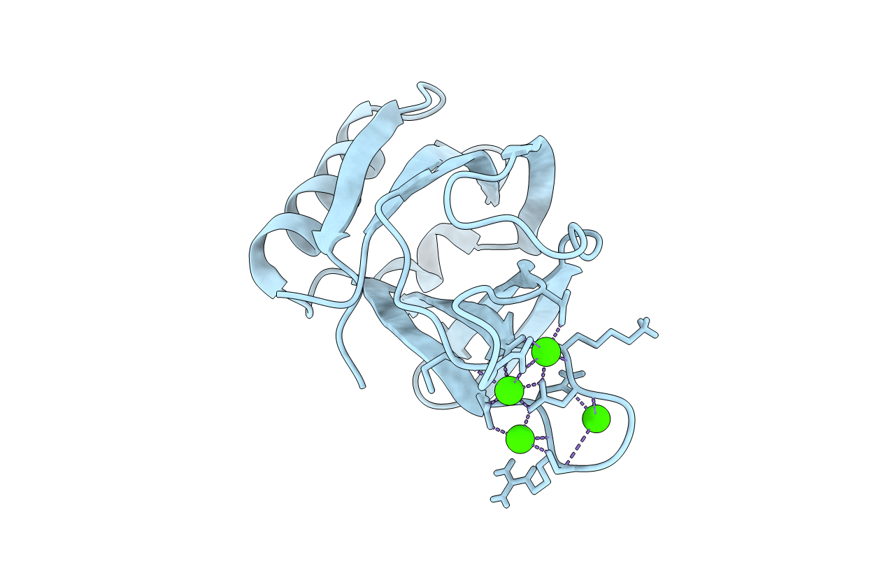
Synaptotagmin-7, C2B-domain, calcium bound
Biological Source:
Source Organism:
Host Organism:
PDB Version:
Deposition Date:
2010-05-24
Release Date:
2010-09-29
Method Details:
Experimental Method:
Resolution:
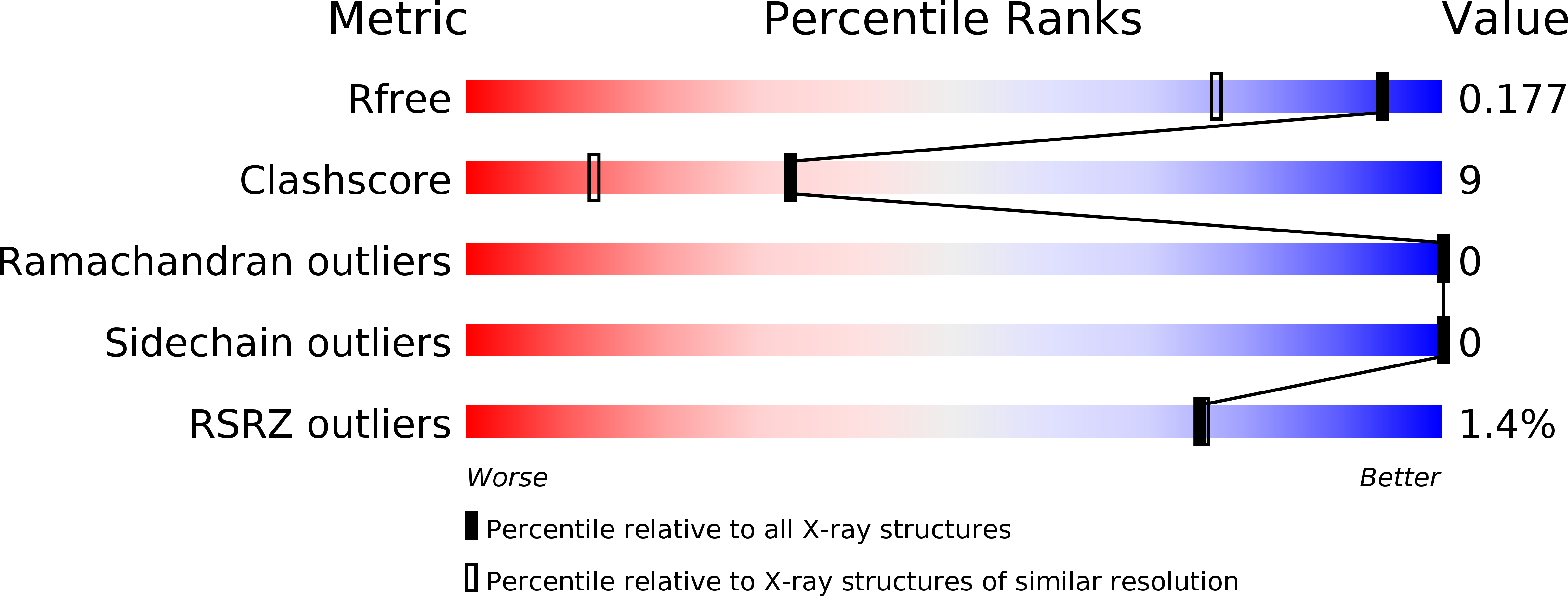
1.44 Å
R-Value Free:
0.18
R-Value Work:
0.16
R-Value Observed:
0.16
Space Group:
P 21 21 21
Macromolecular Entities
Polymer Type:polypeptide(L)
Description:Synaptotagmin-7
Chain IDs:A
Chain Length:138
Number of Molecules:1
Biological Source:Mus musculus
Ligand Molecules
Primary Citation
Structural and mutational analysis of functional differentiation between synaptotagmins-1 and -7.
Plos One 5 12544 12544 (2010)
PMID: 20824061 DOI: 10.1371/journal.pone.0012544

Abstact

Synaptotagmins are known to mediate diverse forms of Ca2+-triggered exocytosis through their C2 domains, but the principles underlying functional differentiation among them are unclear. Synaptotagmin-1 functions as a Ca2+ sensor in neurotransmitter release at central nervous system synapses, but synaptotagmin-7 does not, and yet both isoforms act as Ca2+ sensors in chromaffin cells. To shed light into this apparent paradox, we have performed rescue experiments in neurons from synaptotagmin-1 knockout mice using a chimera that contains the synaptotagmin-1 sequence with its C2B domain replaced by the synaptotagmin-7 C2B domain (Syt1/7). Rescue was not achieved either with the WT Syt1/7 chimera or with nine mutants where residues that are distinct in synaptotagmin-7 were restored to those present in synaptotagmin-1. To investigate whether these results arise because of unique conformational features of the synaptotagmin-7 C2B domain, we determined its crystal structure at 1.44 A resolution. The synaptotagmin-7 C2B domain structure is very similar to that of the synaptotagmin-1 C2B domain and contains three Ca2+-binding sites. Two of the Ca2+-binding sites of the synaptotagmin-7 C2B domain are also present in the synaptotagmin-1 C2B domain and have analogous ligands to those determined for the latter by NMR spectroscopy, suggesting that a discrepancy observed in a crystal structure of the synaptotagmin-1 C2B domain arose from crystal contacts. Overall, our results suggest that functional differentiation in synaptotagmins arises in part from subtle sequence changes that yield dramatic functional differences.

Legend

Protein

Chemical

Disease

Primary Citation of related structures