Deposition Date
2010-05-20
Release Date
2010-06-16
Last Version Date
2024-10-16
Entry Detail
PDB ID:
3N3T
Keywords:
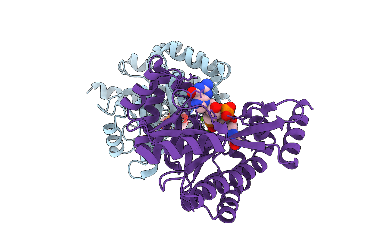
Title:
Crystal structure of putative diguanylate cyclase/phosphodiesterase complex with cyclic di-gmp
Biological Source:
Source Organism(s):
THIOBACILLUS DENITRIFICANS (Taxon ID: 292415)
Expression System(s):
Method Details:
Experimental Method:
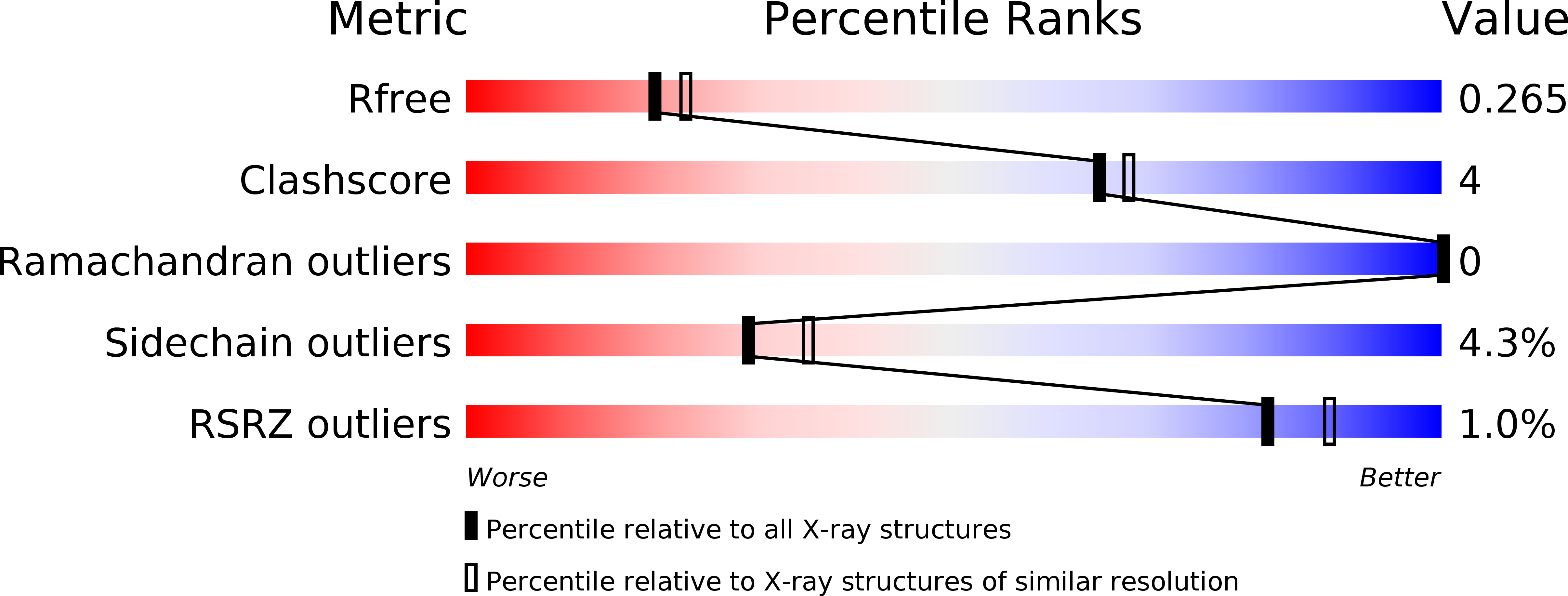
Resolution:
2.35 Å
R-Value Free:
0.25
R-Value Work:
0.18
R-Value Observed:
0.18
Space Group:
P 21 21 21