Deposition Date
2010-05-20
Release Date
2010-06-09
Last Version Date
2024-03-20
Entry Detail
PDB ID:
3N3I
Keywords:
Title:
Crystal Structure of G48V/C95F tethered HIV-1 Protease/Saquinavir complex
Biological Source:
Source Organism(s):
Human immunodeficiency virus type 1 (Taxon ID: 11706)
Expression System(s):
Method Details:
Experimental Method:
Resolution:
2.50 Å
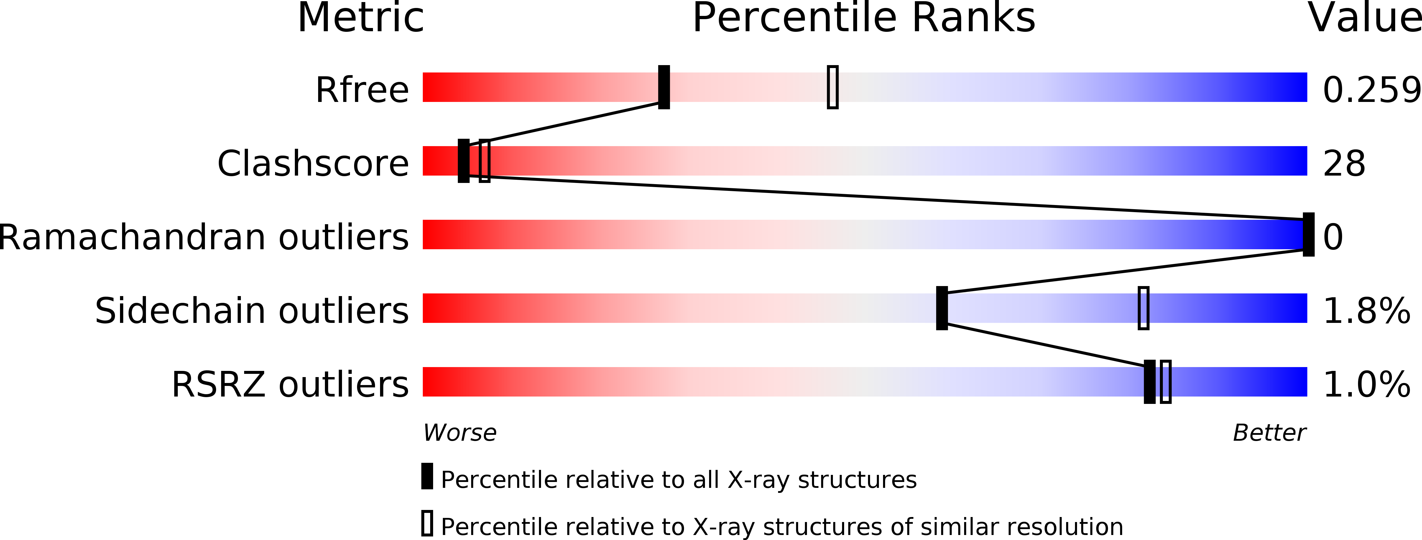
R-Value Free:
0.25
R-Value Work:
0.21
R-Value Observed:
0.21
Space Group:
P 61