3N2T image
Deposition Date 2010-05-19
Release Date 2010-07-21
Last Version Date 2024-02-21
Entry Detail
PDB ID:
3N2T
Keywords:
Title:
Structure of the glycerol dehydrogenase AKR11B4 from Gluconobacter oxydans
Biological Source:
Source Organism:
Host Organism:
Method Details:
Experimental Method:
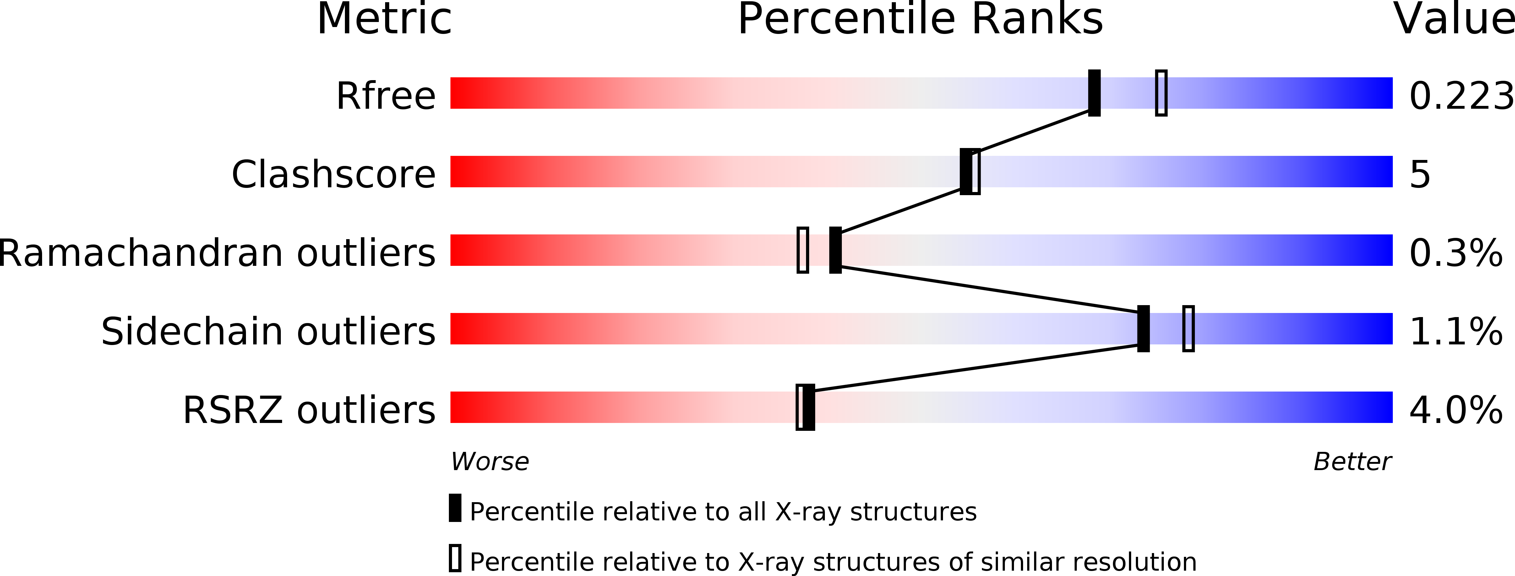
Resolution:
2.00 Å
R-Value Free:
0.23
R-Value Work:
0.18
R-Value Observed:
0.19
Space Group:
P 1 21 1
Macromolecular Entities
Polymer Type:polypeptide(L)
Molecule:Putative oxidoreductase
Gene (Uniprot):GOX1615
Chain IDs:A
Chain Length:348
Number of Molecules:1
Biological Source:Gluconobacter oxydans
Primary Citation
The Three-Dimensional Structure of AKR11B4, a Glycerol Dehydrogenase from Gluconobacter oxydans, Reveals a Tryptophan Residue as an Accelerator of Reaction Turnover.
J.Mol.Biol. 404 353 362 (2010)
PMID: 20887732 DOI: 10.1016/j.jmb.2010.09.049

Abstact

The NADP-dependent glycerol dehydrogenase (EC 1.1.1.72) from Gluconobacter oxydans is a member of family 11 of the aldo-keto reductase (AKR) enzyme superfamily; according to the systematic nomenclature within the AKR superfamily, the term AKR11B4 has been assigned to the enzyme. AKR11B4 is a biotechnologically attractive enzyme because of its broad substrate spectrum, combined with its distinctive regioselectivity and stereoselectivity. These features can be partially rationalized based on a 2-Å crystal structure of apo-AKR11B4, which we describe and interpret here against the functional complex structures of other members of family 11 of the AKR superfamily. The structure of AKR11B4 shows the AKR-typical (β/α)(8) TIM-barrel fold, with three loops and the C-terminal tail determining the particular enzymatic properties. In comparison to AKR11B1 (its closest AKR relative), AKR11B4 has a relatively broad binding cleft for the cosubstrate NADP/NADPH. In the crystalline environment, it is completely blocked by the C-terminal segment of a neighboring protomer. The structure reveals a conspicuous tryptophan residue (Trp23) that has to adopt an unconventional and strained side-chain conformation to permit cosubstrate binding. We predict and confirm by site-directed mutagenesis that Trp23 is an accelerator of (co)substrate turnover. Furthermore, we show that, simultaneously, this tryptophan residue is a critical determinant for substrate binding by the enzyme, while enantioselectivity is probably governed by a methionine residue within the C-terminal tail. We present structural reasons for these notions based on ternary complex models of AKR11B4, NADP, and either octanal, d-glyceraldehyde, or l-glyceraldehyde.

Legend

Protein

Chemical

Disease

Primary Citation of related structures