3N23 image
Deposition Date 2010-05-17
Release Date 2011-01-19
Last Version Date 2025-03-26
Entry Detail
PDB ID:
3N23
Keywords:
Title:
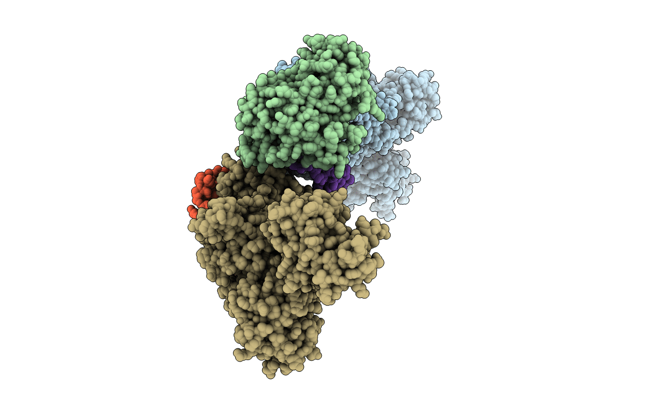
Crystal structure of the high affinity complex between ouabain and the E2P form of the sodium-potassium pump
Biological Source:
Source Organism:
Sus scrofa (Taxon ID: 9823)
Method Details:
Experimental Method:
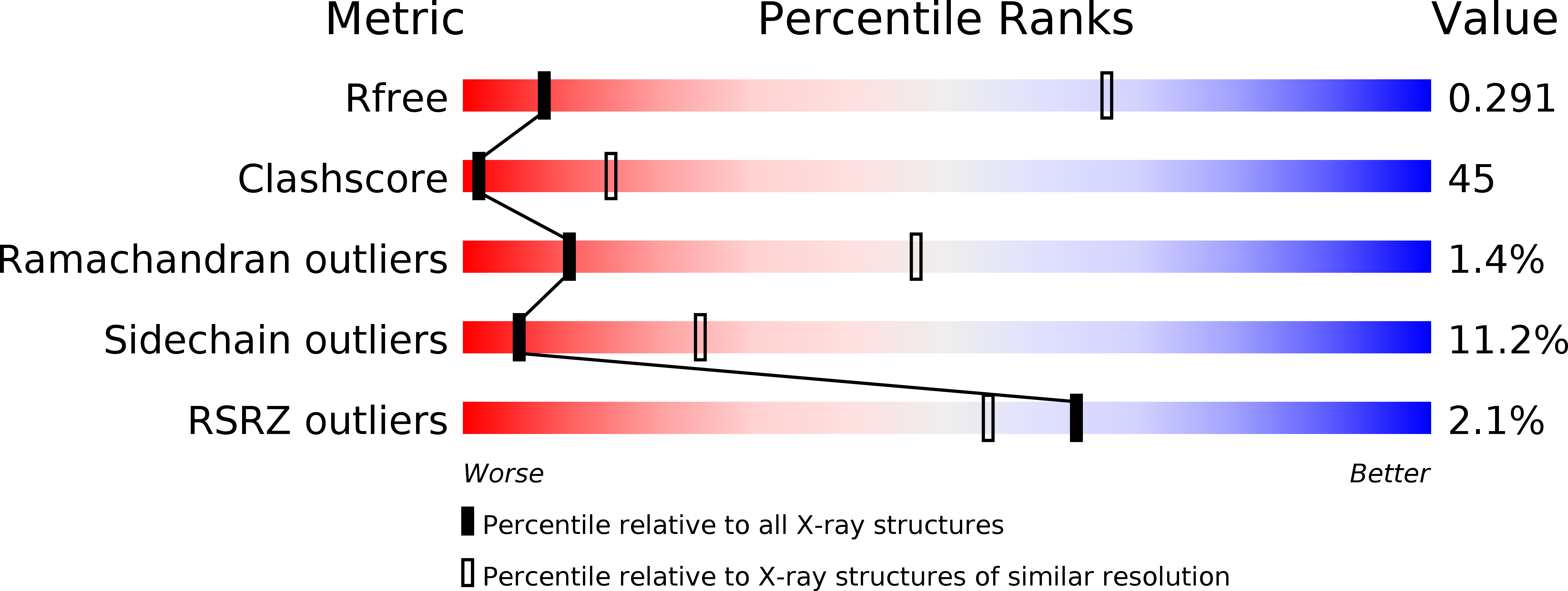
Resolution:
4.60 Å
R-Value Free:
0.30
R-Value Work:
0.27
R-Value Observed:
0.27
Space Group:
P 21 21 21
Macromolecular Entities
Structures with similar UniProt ID
Protein Blast
Polymer Type:polypeptide(L)
Molecule:Sodium/potassium-transporting ATPase subunit alpha-1
Gene (Uniprot):ATP1A1
Chain IDs:A, D (auth: C)
Chain Length:992
Number of Molecules:2
Biological Source:Sus scrofa
Structures with similar UniProt ID
Protein Blast
Polymer Type:polypeptide(L)
Molecule:Sodium/potassium-transporting ATPase subunit beta-1
Gene (Uniprot):ATP1B1
Chain IDs:B, E (auth: D)
Chain Length:277
Number of Molecules:2
Biological Source:Sus scrofa
Structures with similar UniProt ID
Protein Blast
Polymer Type:polypeptide(L)
Molecule:Na+/K+ ATPase gamma subunit transcript variant a
Gene (Uniprot):FXYD2
Chain IDs:C (auth: G), F (auth: E)
Chain Length:31
Number of Molecules:2
Biological Source:Sus scrofa
Modified Residue
Compound ID Chain ID Parent Comp ID Details 2D Image
PHD A ASP ASPARTYL PHOSPHATE
Primary Citation
Structural insights into the high affinity binding of cardiotonic steroids to the Na+,K+-ATPase.
J.Struct.Biol. 174 296 306 (2011)
PMID: 21182963 DOI: 10.1016/j.jsb.2010.12.004

Abstact

The Na+,K+-ATPase belongs to the P-ATPase family, whose characteristic property is the formation of a phosphorylated intermediate. The enzyme is also a defined target for cardiotonic steroids which inhibit its functional activity and initiate intracellular signaling. Here we describe the 4.6 Å resolution crystal structure of the pig kidney Na+,K+-ATPase in its phosphorylated form stabilized by high affinity binding of the cardiotonic steroid ouabain. The steroid binds to a site formed at transmembrane segments αM1-αM6, plugging the ion pathway from the extracellular side. This structure differs from the previously reported low affinity complex with potassium. Most importantly, the A domain has rotated in response to phosphorylation and αM1-2 move towards the ouabain molecule, providing for high affinity interactions and closing the ion pathway from the extracellular side. The observed re-arrangements of the Na+,K+-ATPase stabilized by cardiotonic steroids may affect protein-protein interactions within the intracellular signal transduction networks.

Legend

Protein

Chemical

Disease

Primary Citation of related structures