3MYR image
Entry Detail
PDB ID:
3MYR
Keywords:
Title:
Crystal structure of [NiFe] hydrogenase from Allochromatium vinosum in its Ni-A state
Biological Source:
Source Organism:
PDB Version:
Deposition Date:
2010-05-11
Release Date:
2010-08-04
Method Details:
Experimental Method:
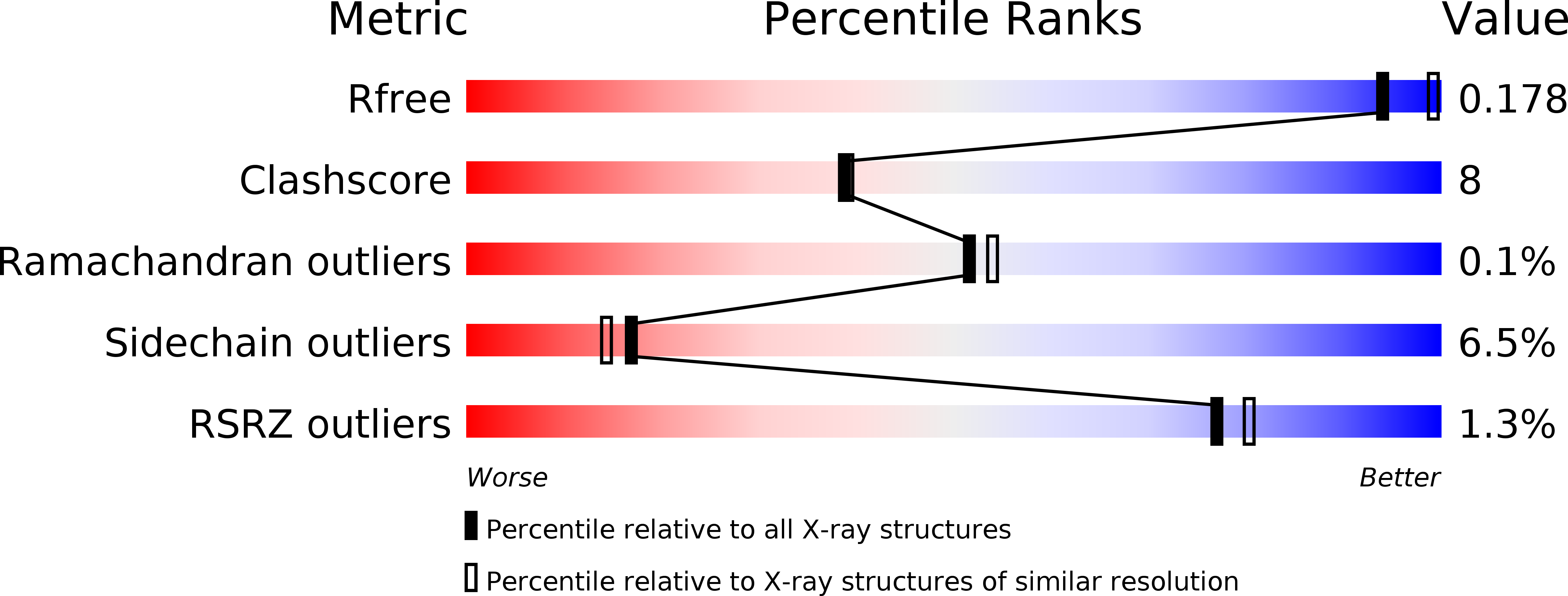
Resolution:
2.10 Å
R-Value Free:
0.16
R-Value Work:
0.13
R-Value Observed:
0.13
Space Group:
P 21 21 2
Macromolecular Entities
Polymer Type:polypeptide(L)
Description:Hydrogenase (NiFe) small subunit HydA
Chain IDs:A, C, E, G
Chain Length:269
Number of Molecules:4
Biological Source:Allochromatium vinosum
Polymer Type:polypeptide(L)
Description:Nickel-dependent hydrogenase large subunit
Chain IDs:B, D, F, H
Chain Length:561
Number of Molecules:4
Biological Source:Allochromatium vinosum
Primary Citation
The crystal structure of the [NiFe] hydrogenase from the photosynthetic bacterium Allochromatium vinosum: characterization of the oxidized enzyme (Ni-A state).
J.Mol.Biol. 402 428 444 (2010)
PMID: 20673834 DOI: 10.1016/j.jmb.2010.07.041

Abstact

The crystal structure of the membrane-associated [NiFe] hydrogenase from Allochromatium vinosum has been determined to 2.1 Å resolution. Electron paramagnetic resonance (EPR) and Fourier transform infrared spectroscopy on dissolved crystals showed that it is present in the Ni-A state (>90%). The structure of the A. vinosum [NiFe] hydrogenase shows significant similarities with [NiFe] hydrogenase structures derived from Desulfovibrio species. The amino acid sequence identity is ∼ 50%. The bimetallic [NiFe] active site is located in the large subunit of the heterodimer and possesses three diatomic non-protein ligands coordinated to the Fe (two CN(-) , one CO). Ni is bound to the protein backbone via four cysteine thiolates; two of them also bridge the two metals. One of the bridging cysteines (Cys64) exhibits a modified thiolate in part of the sample. A mono-oxo bridging ligand was assigned between the metal ions of the catalytic center. This is in contrast to a proposal for Desulfovibrio sp. hydrogenases that show a di-oxo species in this position for the Ni-A state. The additional metal site located in the large subunit appears to be a Mg(2+) ion. Three iron-sulfur clusters were found in the small subunit that forms the electron transfer chain connecting the catalytic site with the molecular surface. The calculated anomalous Fourier map indicates a distorted proximal iron-sulfur cluster in part of the crystals. This altered proximal cluster is supposed to be paramagnetic and is exchange coupled to the Ni(3+) ion and the medial [Fe(3)S(4)](+) cluster that are both EPR active (S=1/2 species). This finding of a modified proximal cluster in the [NiFe] hydrogenase might explain the observation of split EPR signals that are occasionally detected in the oxidized state of membrane-bound [NiFe] hydrogenases as from A. vinosum.

Legend

Protein

Chemical

Disease

Primary Citation of related structures