3MYD image
Deposition Date 2010-05-10
Release Date 2010-05-26
Last Version Date 2024-02-21
Entry Detail
PDB ID:
3MYD
Title:
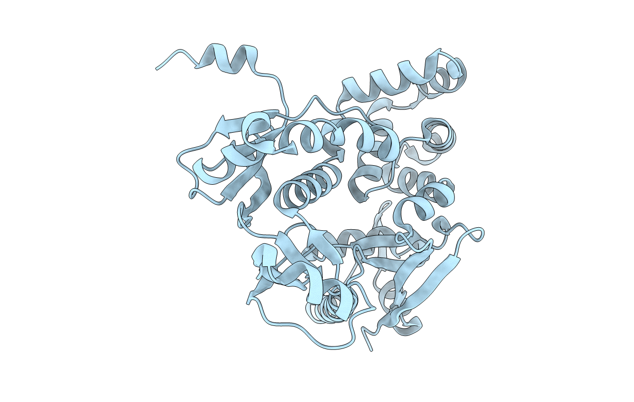
Structure of the Cytoplasmic domain of FlhA from Helicobacter pylori
Biological Source:
Source Organism:
Host Organism:
Method Details:
Experimental Method:
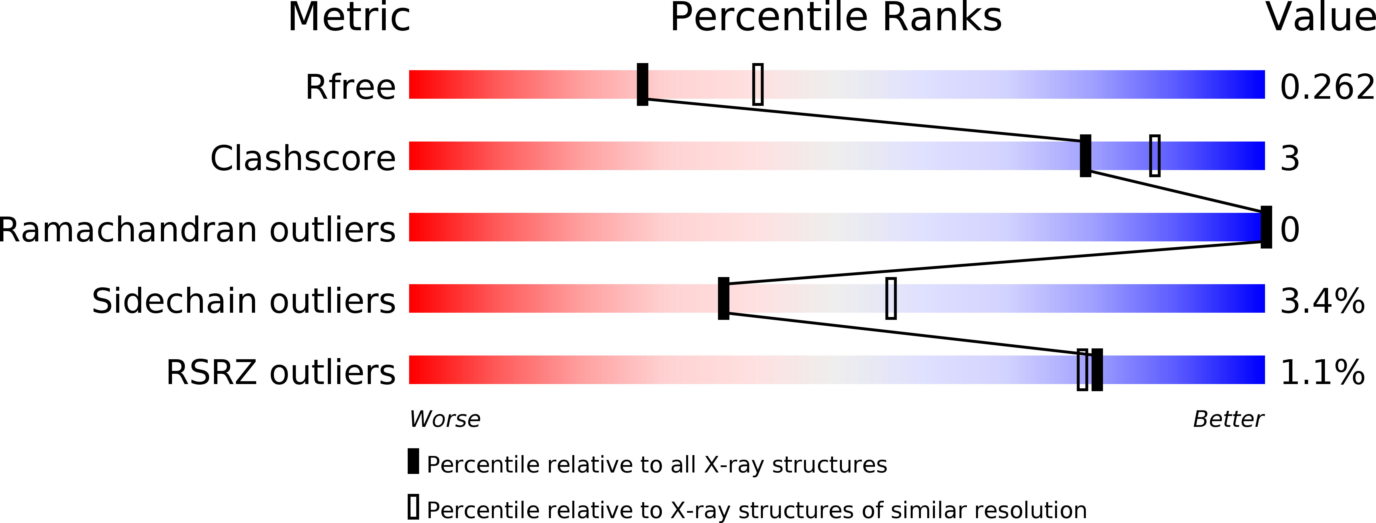
Resolution:
2.40 Å
R-Value Free:
0.26
R-Value Work:
0.22
R-Value Observed:
0.22
Space Group:
C 2 2 21
Macromolecular Entities
Structures with similar UniProt ID
Protein Blast
Polymer Type:polypeptide(L)
Molecule:Flagellar biosynthesis protein flhA
Chain IDs:A
Chain Length:365
Number of Molecules:1
Biological Source:Helicobacter pylori
Primary Citation
Structure of the cytoplasmic domain of the flagellar secretion apparatus component FlhA from Helicobacter pylori.
J.Biol.Chem. 285 21060 21069 (2010)
PMID: 20442410 DOI: 10.1074/jbc.M110.119412

Abstact

Using x-ray crystallography we have determined the structure of the cytoplasmic fragment (residues 384-732) of the flagellum secretion system protein FlhA from Helicobacter pylori at 2.4-A resolution (r = 0.224; R(free) = 0.263). FlhA proteins and their type III secretion homologues contain an N-terminal integral membrane domain (residues 1-350), a linker segment, and a globular C-terminal cytoplasmic region. The tertiary structure of the cytoplasmic fragment contains a thioredoxin-like domain, an RNA recognition motif-like domain inserted within the thioredoxin-fold, a helical domain, and a C-terminal beta/alpha domain. Inter-domain contacts are extensive and the H. pylori FlhA structure appears to be in a closed conformation where the C-terminal domain closes against the RNA recognition motif-fold domain. Highly conserved surface residues in FlhA proteins are concentrated on a narrow surface strip comprising the thioredoxin-like and helical domains, possibly close to the export channel opening. The conformation of the FlhA N-terminal linker segment suggests a likely orientation for the FlhA cytoplasmic fragment relative to the membrane-embedded export pore. Comparison with the recently published structures of the Salmonella FlhA cytoplasmic fragment and its type III secretion counterpart InvA highlight a conformational change where the C-terminal beta/alpha domain in H. pylori FlhA moves 15 A relative to Salmonella FlhA. The conformational change is complex but primarily involves hinge-like movements of the helical and C-terminal domains. Interpretation of previous mutational screens suggest that the C-terminal domain of FlhA(C) plays a regulatory role in substrate class switching in flagellum export.

Legend

Protein

Chemical

Disease

Primary Citation of related structures