Deposition Date
2010-05-07
Release Date
2010-11-10
Last Version Date
2024-03-13
Entry Detail
PDB ID:
3MXE
Keywords:
Title:
Crystal structure of HIV-1 protease inhibitor, KC32 complexed with wild-type protease
Biological Source:
Source Organism(s):
HIV-1 M:B_ARV2/SF2 (Taxon ID: 11685)
Expression System(s):
Method Details:
Experimental Method:
Resolution:
1.85 Å
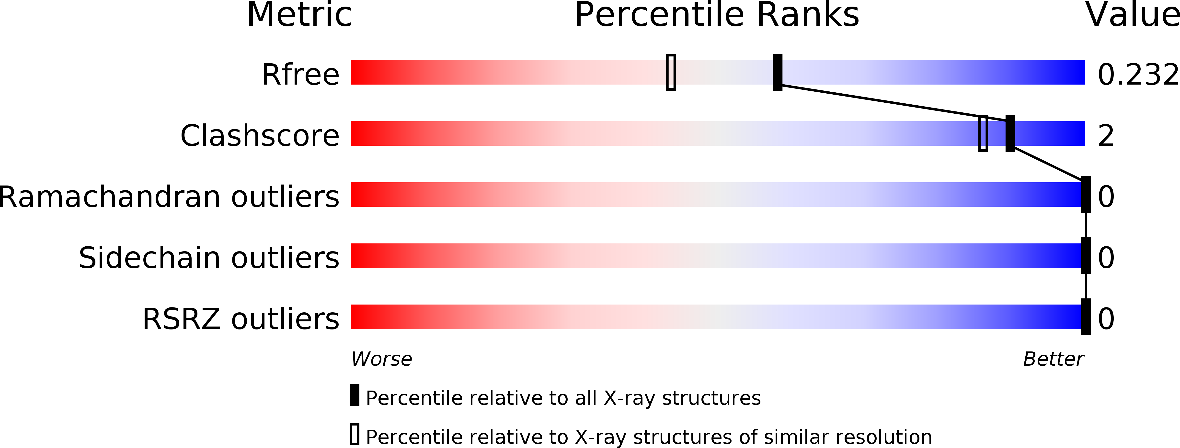
R-Value Free:
0.22
R-Value Work:
0.18
R-Value Observed:
0.18
Space Group:
P 21 21 21