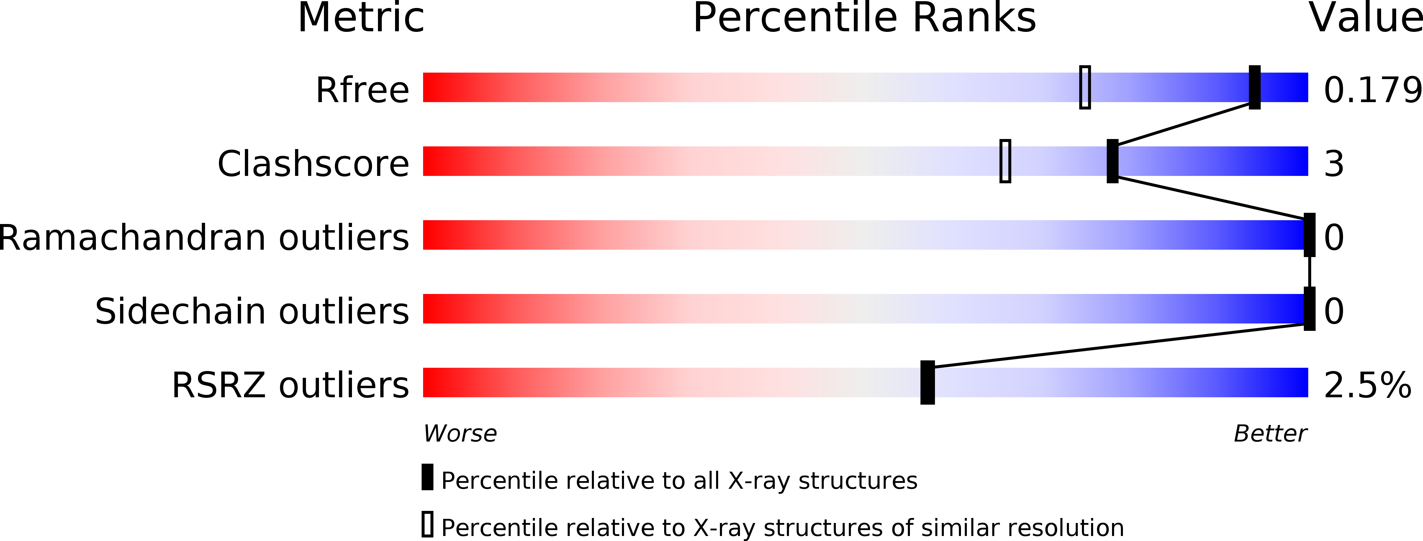
3MWO image
Deposition Date 2010-05-06
Release Date 2010-06-09
Last Version Date 2023-09-06
Entry Detail
PDB ID:
3MWO
Keywords:
Title:
Human carbonic anhydrase II in a doubled monoclinic cell: a re-determination
Biological Source:
Source Organism:
Homo sapiens (Taxon ID: 9606)
Host Organism:
Method Details:
Experimental Method:
Resolution:
1.40 Å
R-Value Free:
0.18
R-Value Work:
0.16
R-Value Observed:
0.16
Space Group:
P 1 21 1
Macromolecular Entities
Polymer Type:polypeptide(L)
Molecule:Carbonic anhydrase 2
Gene (Uniprot):CA2
Chain IDs:A, B
Chain Length:260
Number of Molecules:2
Biological Source:Homo sapiens
Ligand Molecules
Primary Citation
Emerging from pseudo-symmetry: the redetermination of human carbonic anhydrase II in monoclinic P2(1) with a doubled a axis.
Acta Crystallogr.,Sect.D 66 950 952 (2010)
PMID: 20693695 DOI: 10.1107/S0907444910023723

Abstact

The crystal structure of human carbonic anhydrase II in the monoclinic P2(1) space group with a doubled a axis from that of the usually observed unit cell has recently been reported, with one of the two molecules in the asymmetric unit demonstrating rotational disorder [Robbins et al. (2010), Acta Cryst. D66, 628-634]. The structure has been redetermined, with the coordinates of both pseudo-symmetrically related molecules in the crystallographic asymmetric unit translated by x' = x +/- 1/4, and no rotational disorder is observed. This corresponds to a different choice of how the four molecules in the unit cell should be grouped into pairs that represent a single asymmetric unit.

Legend

Protein

Chemical

Disease

Primary Citation of related structures