Deposition Date
2010-05-06
Release Date
2010-06-30
Last Version Date
2024-10-30
Entry Detail
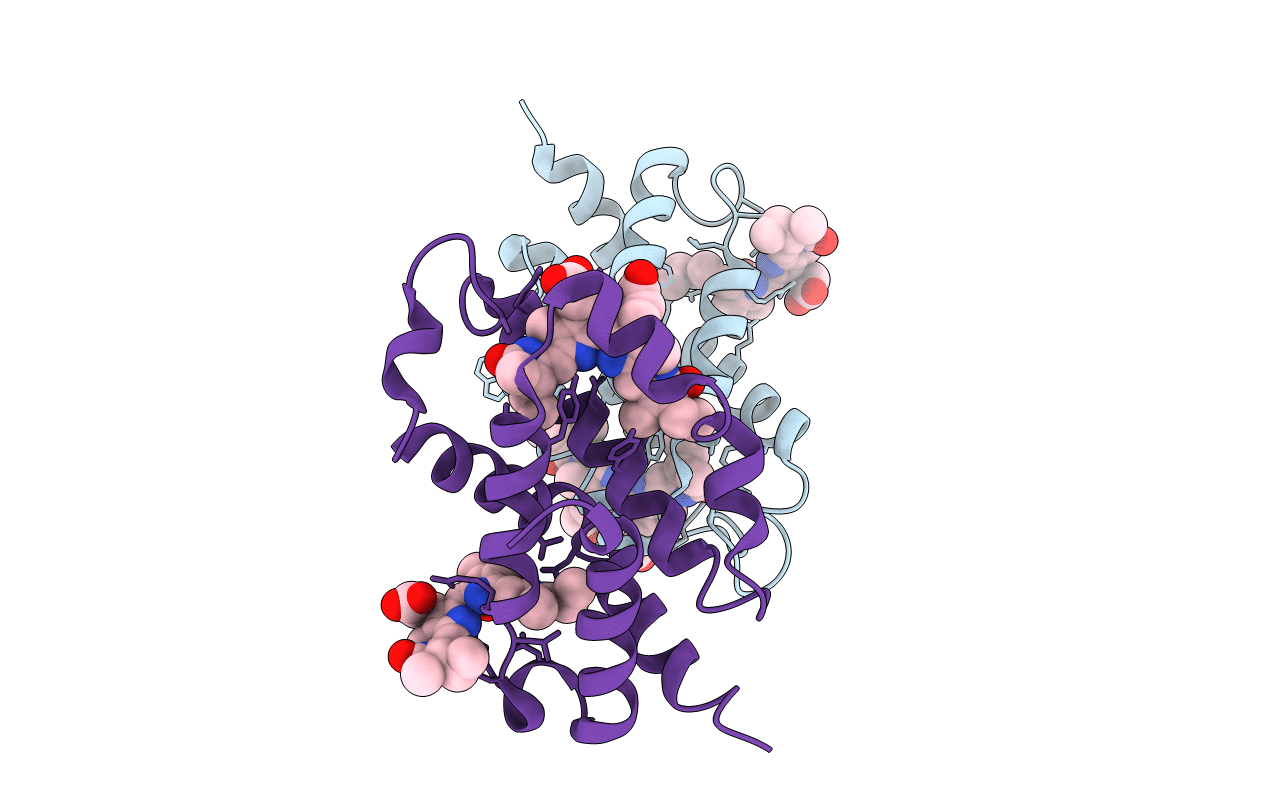
PDB ID:
3MWN
Keywords:
Title:
Structure of the Novel 14 kDa Fragment of alpha-Subunit of Phycoerythrin from the Starving Cyanobacterium Phormidium Tenue
Biological Source:
Source Organism(s):
PHORMIDIUM TENUE (Taxon ID: 126344)
Method Details:
Experimental Method:
Resolution:
2.60 Å
R-Value Free:
0.28
R-Value Work:
0.23
R-Value Observed:
0.23
Space Group:
P 1 21 1