3MT7 image
Deposition Date 2010-04-30
Release Date 2011-01-05
Last Version Date 2023-11-22
Entry Detail
PDB ID:
3MT7
Title:
Glycogen phosphorylase complexed with 4-bromobenzaldehyde-4-(beta-D-glucopyranosyl)-thiosemicarbazone
Biological Source:
Source Organism:
Method Details:
Experimental Method:
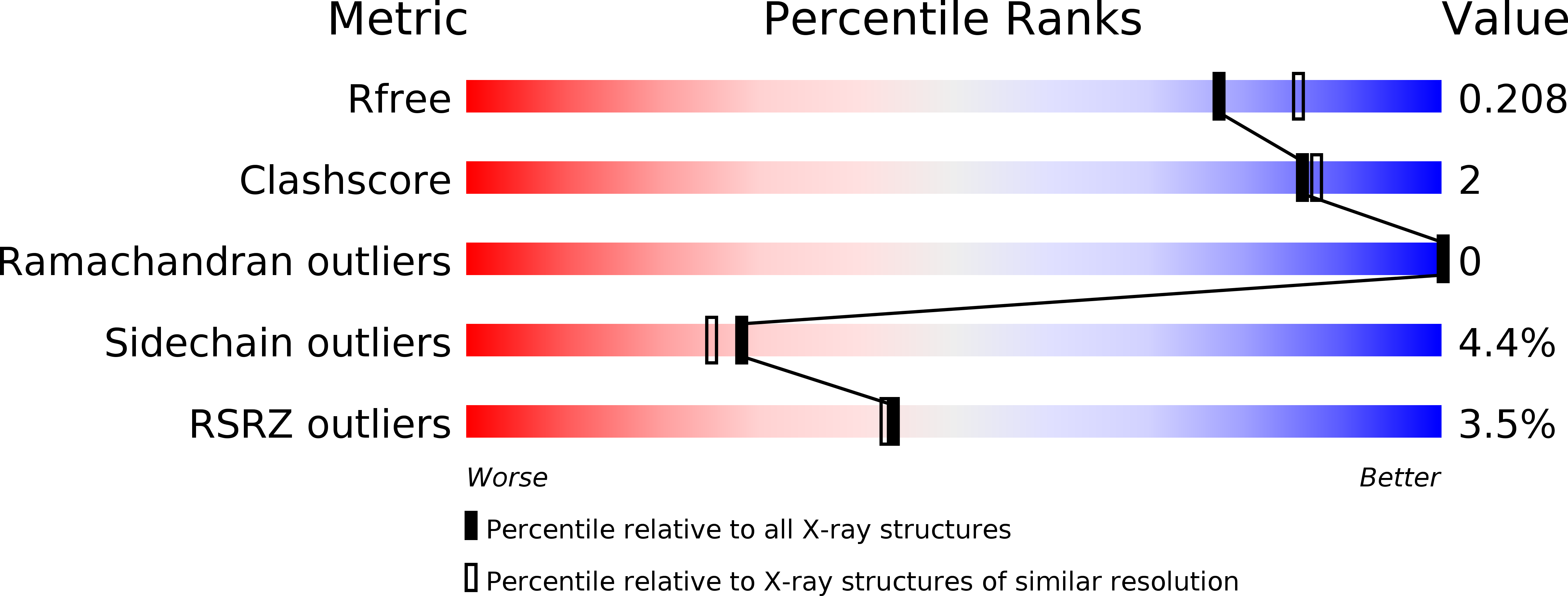
Resolution:
2.00 Å
R-Value Free:
0.20
R-Value Work:
0.17
R-Value Observed:
0.17
Space Group:
P 43 21 2
Macromolecular Entities
Polymer Type:polypeptide(L)
Molecule:Glycogen phosphorylase, muscle form
Gene (Uniprot):PYGM
Chain IDs:A
Chain Length:842
Number of Molecules:1
Biological Source:Oryctolagus cuniculus
Modified Residue
Compound ID Chain ID Parent Comp ID Details 2D Image
LLP A LYS ?
Ligand Molecules
Primary Citation
The binding of beta-D-glucopyranosyl-thiosemicarbazone derivatives to glycogen phosphorylase: a new class of inhibitors
Bioorg.Med.Chem. 18 7911 7922 (2010)
PMID: 20947361 DOI: 10.1016/j.bmc.2010.09.039

Abstact

Glycogen phosphorylase (GP) is a promising target for the treatment of type 2 diabetes. In the process of structure based drug design for GP, a group of 15 aromatic aldehyde 4-(β-d-glucopyranosyl)thiosemicarbazones have been synthesized and evaluated as inhibitors of rabbit muscle glycogen phosphorylase b (GPb) by kinetic studies. These compounds are competitive inhibitors of GPb with respect to α-d-glucose-1-phosphate with IC(50) values ranging from 5.7 to 524.3μM. In order to elucidate the structural basis of their inhibition, the crystal structures of these compounds in complex with GPb at 1.95-2.23Å resolution were determined. The complex structures reveal that the inhibitors are accommodated at the catalytic site with the glucopyranosyl moiety at approximately the same position as α-d-glucose and stabilize the T conformation of the 280s loop. The thiosemicarbazone part of the studied glucosyl thiosemicarbazones possess a moiety derived from substituted benzaldehydes with NO(2), F, Cl, Br, OH, OMe, CF(3), or Me at the ortho-, meta- or para-position of the aromatic ring as well as a moiety derived from 4-pyridinecarboxaldehyde. These fit tightly into the β-pocket, a side channel from the catalytic site with no access to the bulk solvent. The differences in their inhibitory potency can be interpreted in terms of variations in the interactions of the aldehyde-derived moiety with protein residues in the β-pocket. In addition, 14 out of the 15 studied inhibitors were found bound at the new allosteric site of the enzyme.

Legend

Protein

Chemical

Disease

Primary Citation of related structures