3MPU image
Deposition Date 2010-04-27
Release Date 2010-07-21
Last Version Date 2024-11-20
Entry Detail
PDB ID:
3MPU
Keywords:
Title:
Crystal structure of the C47A/A241C disulfide-linked E. coli Aspartate Transcarbamoylase holoenzyme
Biological Source:
Source Organism:
Host Organism:
Method Details:
Experimental Method:
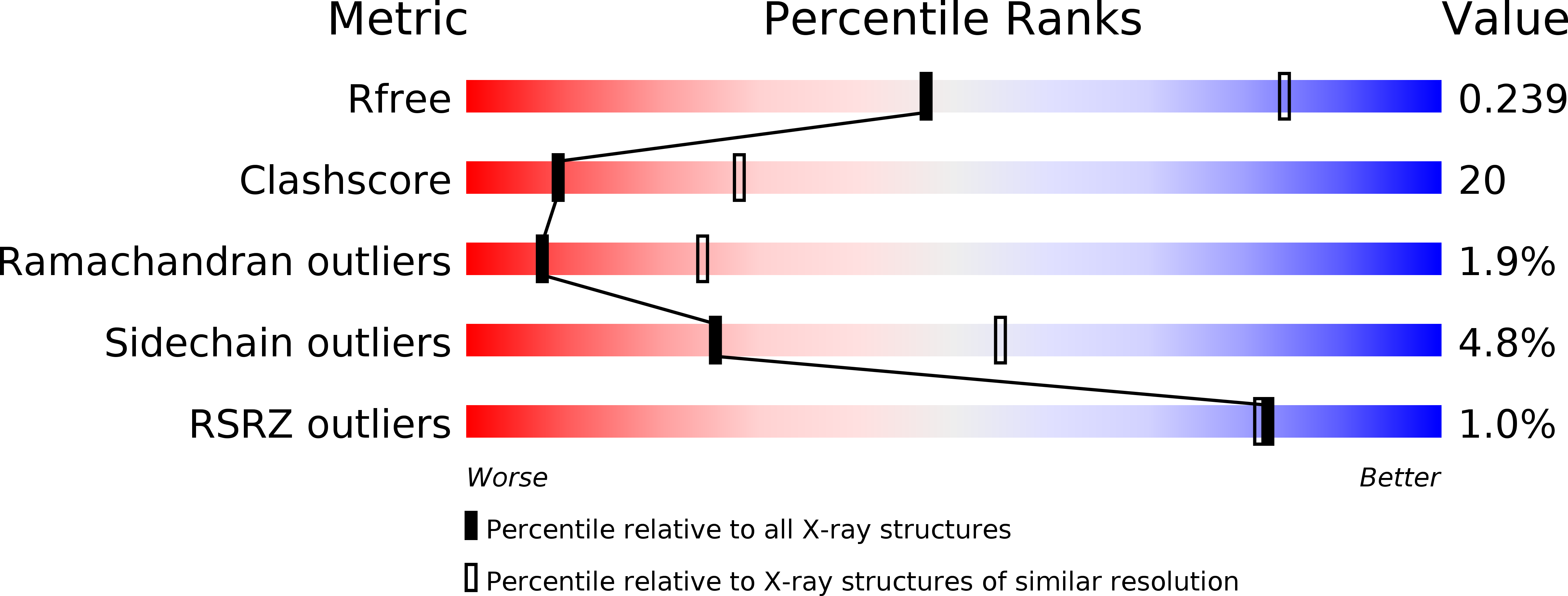
Resolution:
2.86 Å
R-Value Free:
0.23
R-Value Work:
0.17
R-Value Observed:
0.17
Space Group:
H 3 2
Macromolecular Entities
Structures with similar UniProt ID
Protein Blast
Polymer Type:polypeptide(L)
Molecule:Aspartate carbamoyltransferase catalytic chain
Gene (Uniprot):pyrB
Mutagens:C47A, A241C
Chain IDs:A, C, E
Chain Length:310
Number of Molecules:3
Biological Source:Escherichia coli
Structures with similar UniProt ID
Protein Blast
Polymer Type:polypeptide(L)
Molecule:Aspartate carbamoyltransferase regulatory chain
Gene (Uniprot):pyrI
Chain IDs:B, D, F
Chain Length:153
Number of Molecules:3
Biological Source:Escherichia coli
Primary Citation
The Pathway of Product Release from the R State of Aspartate Transcarbamoylase.
J.Mol.Biol. 401 940 948 (2010)
PMID: 20620149 DOI: 10.1016/j.jmb.2010.07.003

Abstact

The pathway of product release from the R state of aspartate transcarbamoylase (ATCase; EC 2.1.3.2, aspartate carbamoyltransferase) has been determined here by solving the crystal structure of Escherichia coli ATCase locked in the R quaternary structure by specific introduction of disulfide bonds. ATCase displays ordered substrate binding and product release, remaining in the R state until substrates are exhausted. The structure reported here represents ATCase in the R state bound to the final product molecule, phosphate. This structure has been difficult to obtain previously because the enzyme relaxes back to the T state after the substrates are exhausted. Hence, cocrystallizing the wild-type enzyme with phosphate results in a T-state structure. In this structure of the enzyme trapped in the R state with specific disulfide bonds, we observe two phosphate molecules per active site. The position of the first phosphate corresponds to the position of the phosphate of carbamoyl phosphate (CP) and the position of the phosphonate of N-phosphonacetyl-l-aspartate. However, the second, more weakly bound phosphate is bound in a positively charged pocket that is more accessible to the surface than the other phosphate. The second phosphate appears to be on the path that phosphate would have to take to exit the active site. Our results suggest that phosphate dissociation and CP binding can occur simultaneously and that the dissociation of phosphate may actually promote the binding of CP for more efficient catalysis.

Legend

Protein

Chemical

Disease

Primary Citation of related structures
Feedback Form
Name
Email
Institute
Feedback