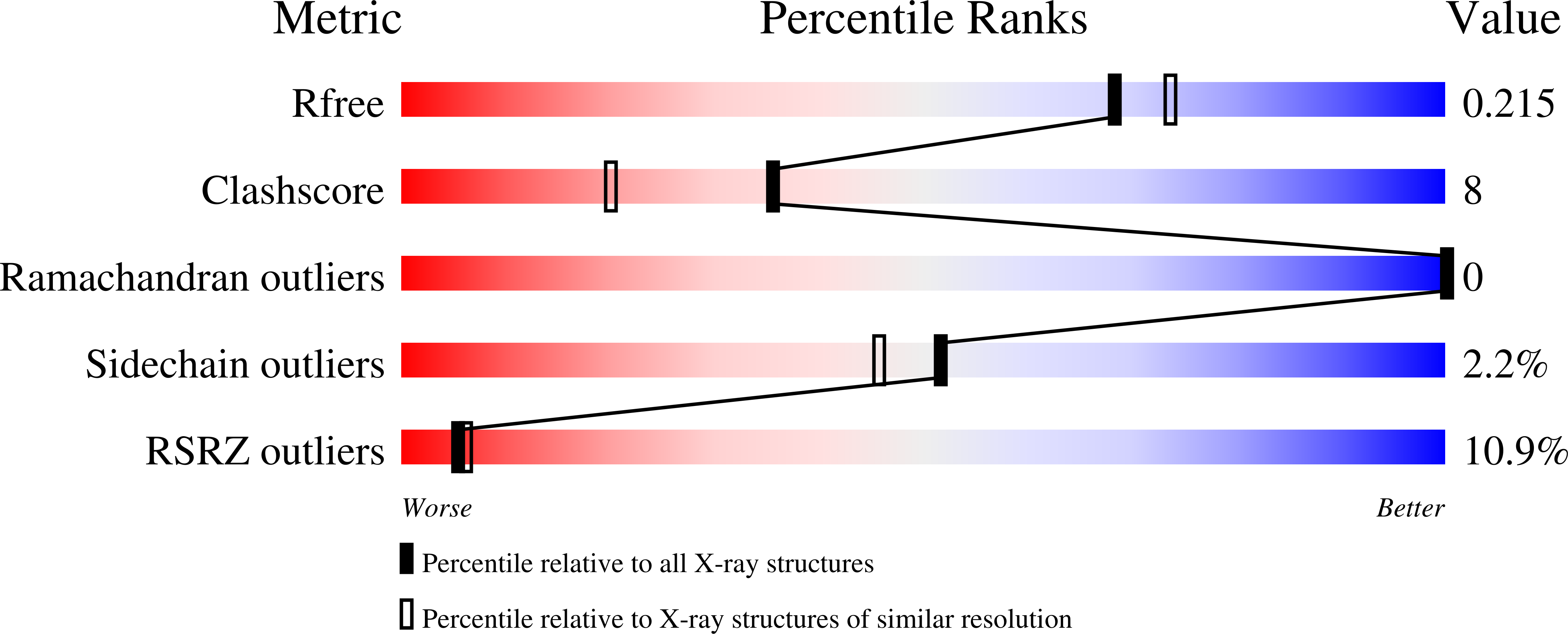
3MPP image
Deposition Date 2010-04-27
Release Date 2010-06-16
Last Version Date 2023-09-06
Entry Detail
PDB ID:
3MPP
Keywords:
Title:
Botulinum Neurotoxin Type G Receptor Binding Domain
Biological Source:
Source Organism:
Host Organism:
Method Details:
Experimental Method:
Resolution:
1.98 Å
R-Value Free:
0.22
R-Value Work:
0.17
R-Value Observed:
0.17
Space Group:
P 21 21 21
Macromolecular Entities
Polymer Type:polypeptide(L)
Molecule:Botulinum neurotoxin type G
Gene (Uniprot):botG
Chain IDs:A (auth: G)
Chain Length:433
Number of Molecules:1
Biological Source:Clostridium botulinum
Primary Citation
Structural analysis of botulinum neurotoxin type G receptor binding .
Biochemistry 49 5200 5205 (2010)
PMID: 20507178 DOI: 10.1021/bi100412v

Abstact

Botulinum neurotoxin (BoNT) binds peripheral neurons at the neuromuscular junction through a dual-receptor mechanism that includes interactions with ganglioside and protein receptors. The receptor identities vary depending on BoNT serotype (A-G). BoNT/B and BoNT/G bind the luminal domains of synaptotagmin I and II, homologous synaptic vesicle proteins. We observe conditions under which BoNT/B binds both Syt isoforms, but BoNT/G binds only SytI. Both serotypes bind ganglioside G(T1b). The BoNT/G receptor-binding domain crystal structure provides a context for examining these binding interactions and a platform for understanding the physiological relevance of different Syt receptor isoforms in vivo.

Legend

Protein

Chemical

Disease

Primary Citation of related structures