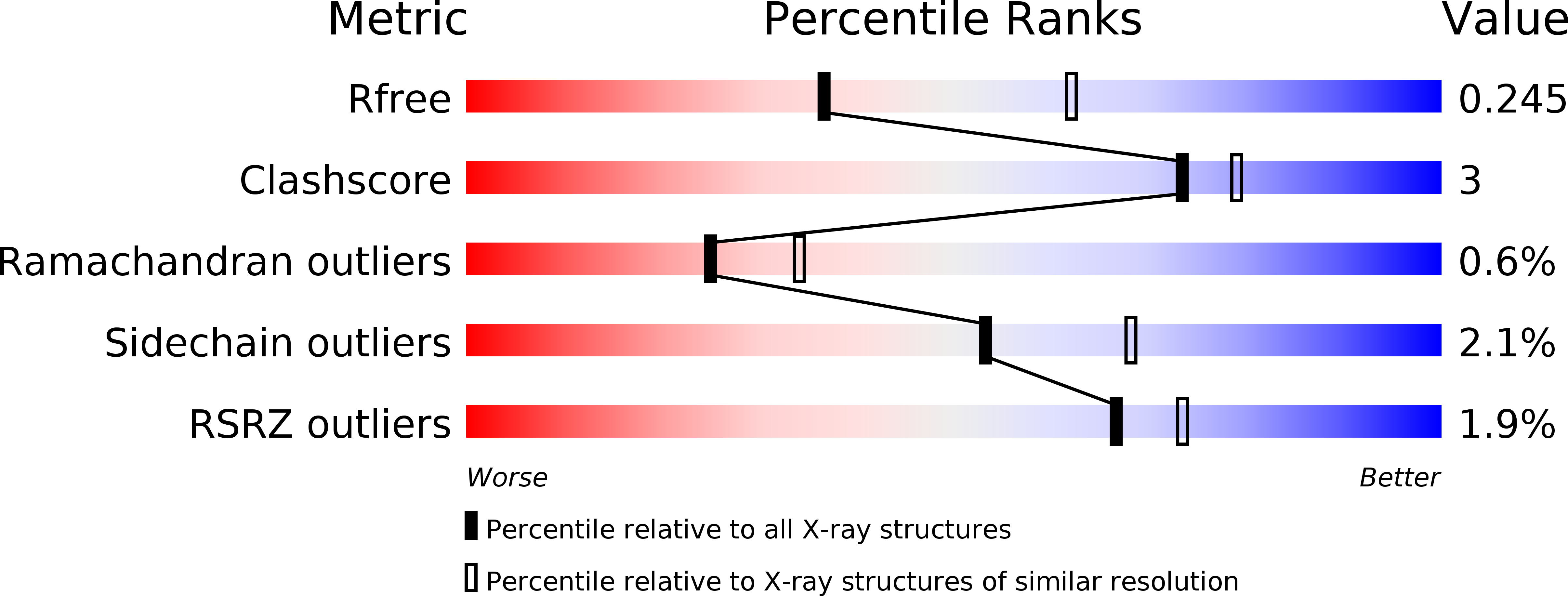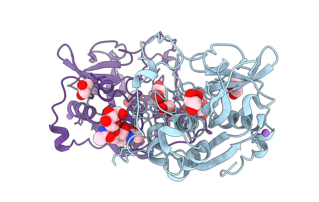
3MOR image
Deposition Date 2010-04-23
Release Date 2011-11-02
Last Version Date 2024-11-06
Entry Detail
PDB ID:
3MOR
Keywords:
Title:
Crystal structure of Cathepsin B from Trypanosoma Brucei
Biological Source:
Source Organism:
Host Organism:
Method Details:
Experimental Method:
Resolution:
2.55 Å
R-Value Free:
0.24
R-Value Work:
0.20
R-Value Observed:
0.20
Space Group:
P 1 21 1
Macromolecular Entities
Polymer Type:polypeptide(L)
Molecule:Cathepsin B-like cysteine protease
Chain IDs:A, B
Chain Length:317
Number of Molecules:2
Biological Source:Trypanosoma brucei
Modified Residue
Compound ID Chain ID Parent Comp ID Details 2D Image
ASN B ASN GLYCOSYLATION SITE
Primary Citation

Abstact

Protein crystallization in cells has been observed several times in nature. However, owing to their small size these crystals have not yet been used for X-ray crystallographic analysis. We prepared nano-sized in vivo-grown crystals of Trypanosoma brucei enzymes and applied the emerging method of free-electron laser-based serial femtosecond crystallography to record interpretable diffraction data. This combined approach will open new opportunities in structural systems biology.

Legend

Protein

Chemical

Disease

Primary Citation of related structures