Deposition Date
2010-04-17
Release Date
2010-12-01
Last Version Date
2024-03-20
Entry Detail
PDB ID:
3MLO
Keywords:
Title:
DNA binding domain of Early B-cell Factor 1 (Ebf1) bound to DNA (Crystal form I)
Biological Source:
Source Organism(s):
Mus musculus (Taxon ID: 10090)
synthetic construct (Taxon ID: 32630)
synthetic construct (Taxon ID: 32630)
Expression System(s):
Method Details:
Experimental Method:
Resolution:
3.01 Å
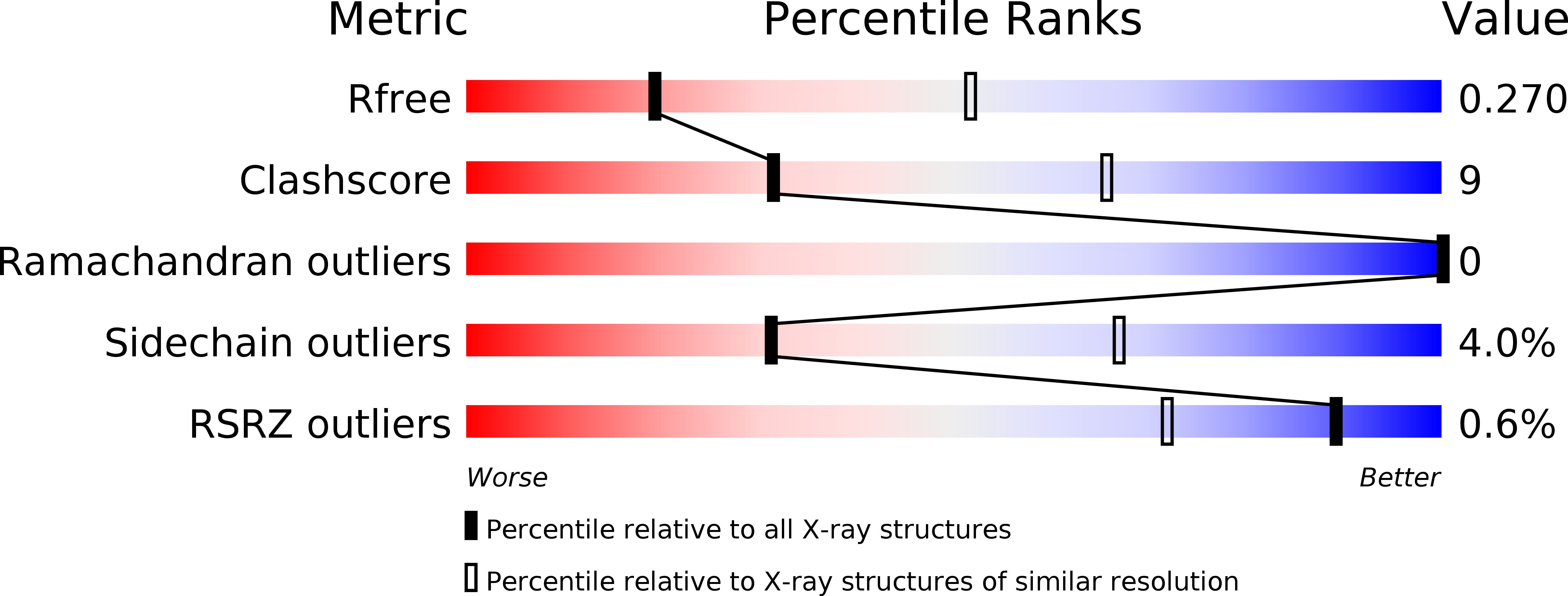
R-Value Free:
0.27
R-Value Work:
0.22
R-Value Observed:
0.22
Space Group:
P 65 2 2