3ML1 image
Entry Detail
PDB ID:
3ML1
Keywords:
Title:
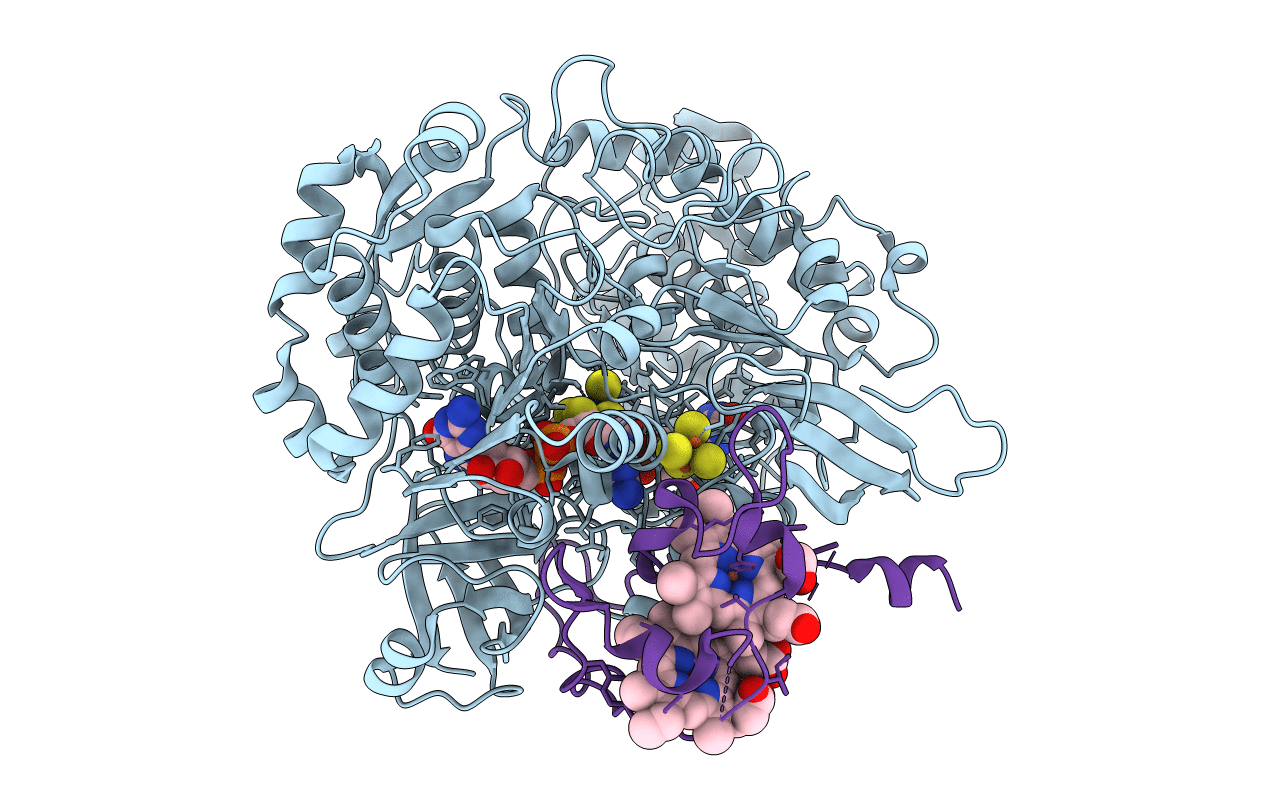
Crystal Structure of the Periplasmic Nitrate Reductase from Cupriavidus necator
Biological Source:
Source Organism:
Host Organism:
PDB Version:
Deposition Date:
2010-04-16
Release Date:
2011-04-06
Method Details:
Experimental Method:
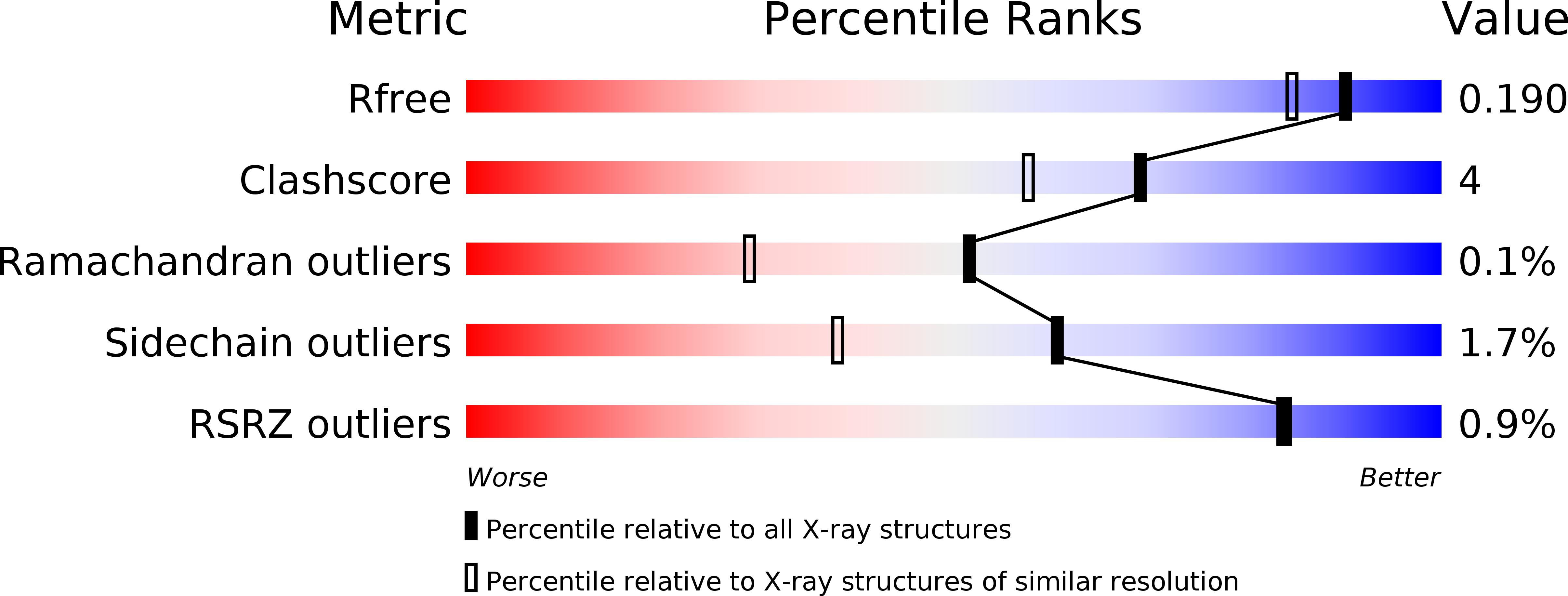
Resolution:
1.60 Å
R-Value Free:
0.19
R-Value Work:
0.15
R-Value Observed:
0.15
Space Group:
C 1 2 1
Macromolecular Entities
Polymer Type:polypeptide(L)
Description:Periplasmic nitrate reductase
Chain IDs:A
Chain Length:802
Number of Molecules:1
Biological Source:Ralstonia eutropha
Polymer Type:polypeptide(L)
Description:Diheme cytochrome c napB
Chain IDs:B
Chain Length:135
Number of Molecules:1
Biological Source:Ralstonia eutropha
Primary Citation
The crystal structure of Cupriavidus necator nitrate reductase in oxidized and partially reduced states
J.Mol.Biol. 408 932 948 (2011)
PMID: 21419779 DOI: 10.1016/j.jmb.2011.03.016

Abstact

The periplasmic nitrate reductase (NapAB) from Cupriavidus necator is a heterodimeric protein that belongs to the dimethyl sulfoxide reductase family of mononuclear Mo-containing enzymes and catalyzes the reduction of nitrate to nitrite. The protein comprises a large catalytic subunit (NapA, 91 kDa) containing the molybdenum active site plus one [4Fe-4S] cluster, as well as a small subunit (NapB, 17 kDa), which is a diheme c-type cytochrome involved in electron transfer. Crystals of the oxidized form of the enzyme diffracted beyond 1.5 Å at the European Synchrotron Radiation Facility. This is the highest resolution reported to date for a nitrate reductase, providing true atomic details of the protein active center, and this showed further evidence on the molybdenum coordination sphere, corroborating previous data on the related Desulfovibrio desulfuricans NapA. The molybdenum atom is bound to a total of six sulfur atoms, with no oxygen ligands or water molecules in the vicinity. In the present work, we were also able to prepare partially reduced crystals that revealed two alternate conformations of the Mo-coordinating cysteine. This crystal form was obtained by soaking dithionite into crystals grown in the presence of the ionic liquid [C(4)mim]Cl(-). In addition, UV-Vis and EPR spectroscopy studies showed that the periplasmic nitrate reductase from C. necator might work at unexpectedly high redox potentials when compared to all periplasmic nitrate reductases studied to date.

Legend

Protein

Chemical

Disease

Primary Citation of related structures