3MKJ image
Deposition Date 2010-04-15
Release Date 2010-08-11
Last Version Date 2024-10-16
Entry Detail
PDB ID:
3MKJ
Keywords:
Title:
Methionine gamma-lyase from Citrobacter freundii with pyridoximine-5'-phosphate
Biological Source:
Source Organism:
Host Organism:
Method Details:
Experimental Method:
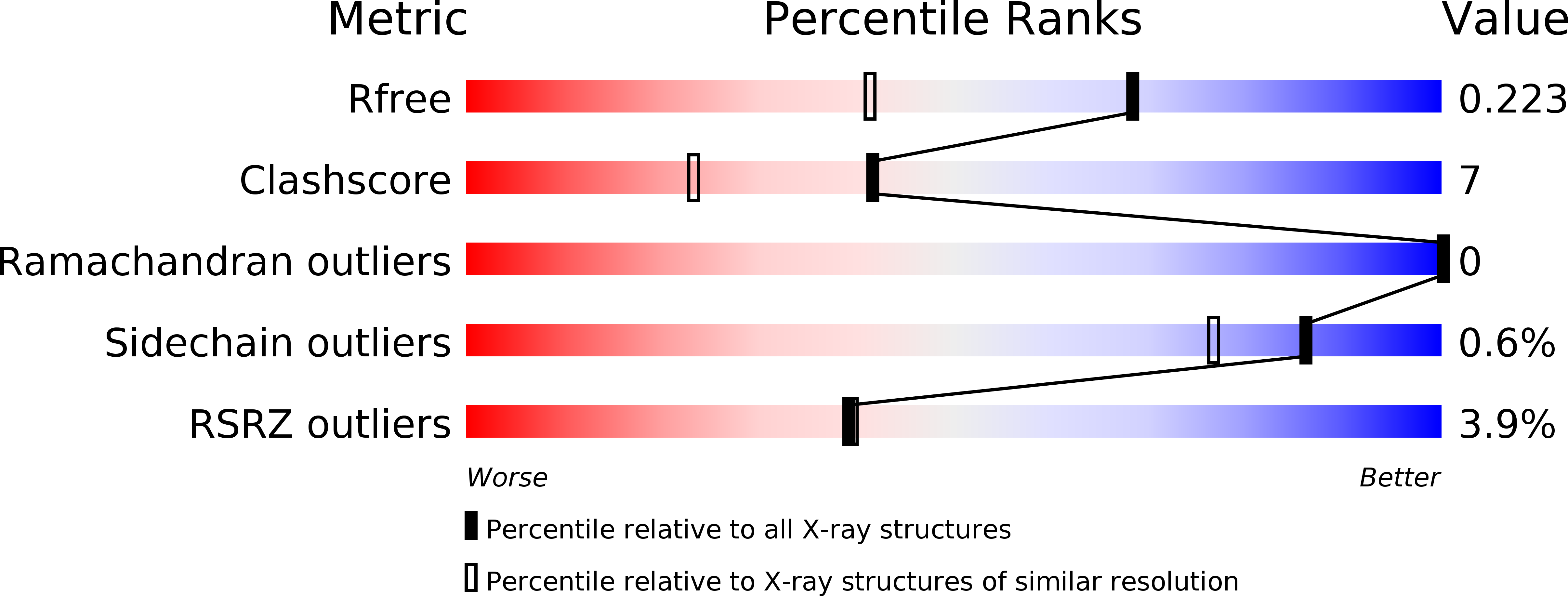
Resolution:
1.65 Å
R-Value Free:
0.23
R-Value Work:
0.19
R-Value Observed:
0.19
Space Group:
I 2 2 2
Macromolecular Entities
Structures with similar UniProt ID
Protein Blast
Polymer Type:polypeptide(L)
Molecule:Methionine gamma-lyase
Gene (Uniprot):megL
Chain IDs:A
Chain Length:398
Number of Molecules:1
Biological Source:Citrobacter freundii
Modified Residue
Compound ID Chain ID Parent Comp ID Details 2D Image
CSO A CYS S-HYDROXYCYSTEINE
Ligand Molecules
Primary Citation
Exploring methionine gamma-lyase structure-function relationship via microspectrophotometry and X-ray crystallography
Biochim.Biophys.Acta 1814 834 842 (2011)
PMID: 20601224 DOI: 10.1016/j.bbapap.2010.06.017

Abstact

Pyridoxal 5'-phosphate (PLP) dependent methionine γ-lyase catalyzes the breakdown of L-methionine to α-ketobutyric acid, methanethiol and ammonia. This enzyme, present in anaerobic microorganisms, has biomedical interest both for its activity as antitumor agent, depleting methionine supply in methionine-dependent cancers, and as target in the treatment of human pathogen infections, activating the pro-drug trifluoromethionine. To validate the structure of the enzyme from Citrobacter freundii, crystallized from monomethyl ether polyethylene glycol 2000, for the development of lead compounds, the reactivity of the crystalline enzyme towards L-methionine, substrate analogs and inhibitors was determined by polarized absorption microspectrophotometry. Spectral data were also collected for enzyme crystals, grown in monomethyl ether polyethylene glycol 2000 in the presence of ammonium sulfate. The three-dimensional structure of these enzyme crystals, solved at 1.65Å resolution with R(free) 23.2%, revealed the surprising absence of the aldimine bond between the active site Lys210 and PLP. Different hypothesis are proposed and discussed in the light of spectral and structural data, pointing out to the relevance of the complementarity between X-ray crystallography and single crystal spectroscopy for the understanding of biological mechanisms at molecular level. This article is part of a Special Issue entitled: Protein Structure and Function in the Crystalline State.

Legend

Protein

Chemical

Disease

Primary Citation of related structures