3MK3 image
Deposition Date 2010-04-14
Release Date 2011-02-02
Last Version Date 2023-11-01
Entry Detail
PDB ID:
3MK3
Keywords:
Title:
Crystal structure of Lumazine synthase from Salmonella typhimurium LT2
Biological Source:
Source Organism:
Host Organism:
Method Details:
Experimental Method:
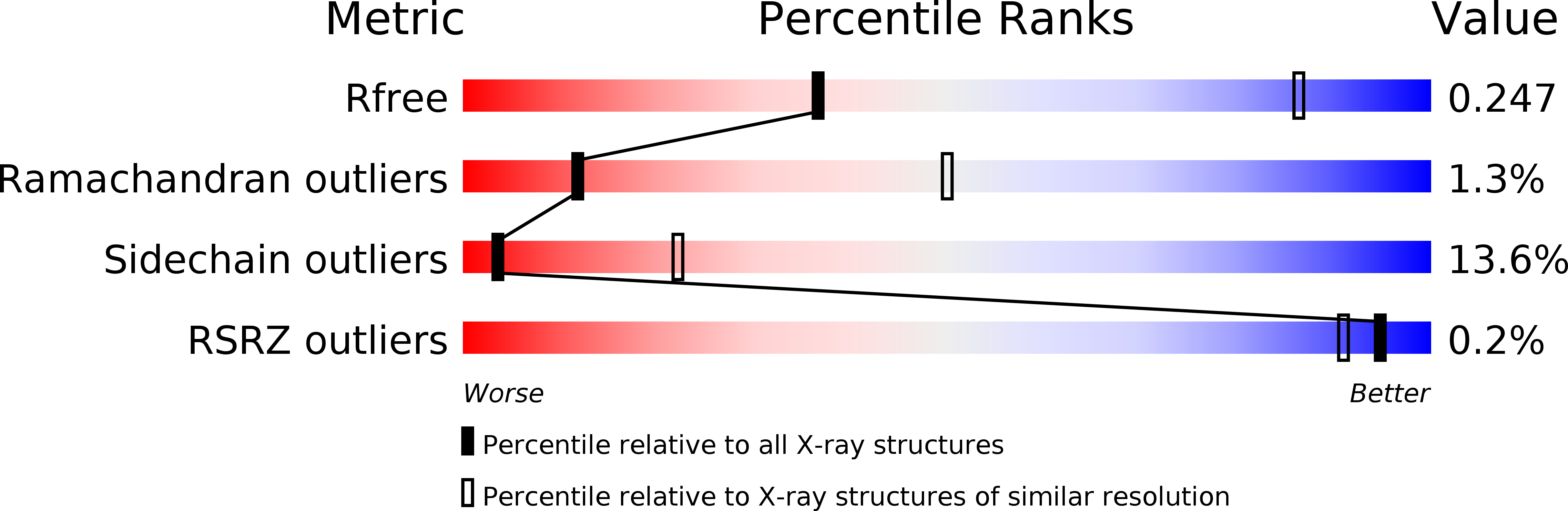
Resolution:
3.57 Å
R-Value Free:
0.26
R-Value Work:
0.22
R-Value Observed:
0.22
Space Group:
P 1 21 1
Macromolecular Entities
Structures with similar UniProt ID
Protein Blast
Polymer Type:polypeptide(L)
Molecule:6,7-dimethyl-8-ribityllumazine synthase
Gene (Uniprot):ribH
Chain IDs:A, B, C, D, E, F, G, H, I, J, K, L, M, N, O, P, Q, R, S, T, U, V, W, X, Y, Z, AA (auth: 1), BA (auth: 2), CA (auth: 3), DA (auth: 4), EA (auth: 5), FA (auth: 6), GA (auth: 7), HA (auth: 8), IA (auth: 9), JA (auth: a), KA (auth: b), LA (auth: c), MA (auth: d), NA (auth: e), OA (auth: f), PA (auth: g), QA (auth: h), RA (auth: i), SA (auth: j), TA (auth: k), UA (auth: l), VA (auth: m), WA (auth: n), XA (auth: o), YA (auth: p), ZA (auth: q), AB (auth: r), BB (auth: s), CB (auth: t), DB (auth: u), EB (auth: v), FB (auth: w), GB (auth: x), HB (auth: y)
Chain Length:156
Number of Molecules:60
Biological Source:Salmonella typhimurium
Ligand Molecules
Primary Citation
Crystal structure analysis of icosahedral lumazine synthase from Salmonella typhimurium, an antibacterial drug target.
Acta Crystallogr.,Sect.D 67 131 139 (2011)
PMID: 21245535 DOI: 10.1107/S0907444910053370

Abstact

Riboflavin biosynthesis is an essential pathway in bacteria, in contrast to animals, which obtain riboflavin from their diet. Therefore, the enzymes involved in the riboflavin-biosynthesis pathway are potential targets for the development of antibacterial drugs. Lumazine synthase, an enzyme that is involved in the penultimate step of riboflavin biosynthesis, catalyzes the formation of 6,7-dimethyl-8-ribityllumazine from 3,4-dihydroxy-2-butanone 4-phosphate and 5-amino-6-ribitylamino-2,4-(1H,3H)-pyrimidinedione. Lumazine synthase from Salmonella typhimurium (sLS) has been cloned, overexpressed, purified and was crystallized in three forms, each with different crystal packing. The crystal structure of sLS in the monoclinic space group P2(1) has been determined with 60 subunits per asymmetric unit, packed as an icosahedron, at 3.57 Å resolution. Interestingly, sLS contains an N-terminal proline residue (Pro11) which had previously been suggested to disrupt the formation of the icosohedral assembly. In addition, comparison of the structure of sLS with known orthologous lumazine synthase structures allowed identification of the amino-acid residues involved in substrate binding and catalysis. The sLS structure reported here could serve as a starting point for the development of species-specific antibacterial drugs.

Legend

Protein

Chemical

Disease

Primary Citation of related structures
Feedback Form
Name
Email
Institute
Feedback