3MIO image
Deposition Date 2010-04-11
Release Date 2011-02-23
Last Version Date 2023-11-01
Entry Detail
PDB ID:
3MIO
Keywords:
Title:
Crystal structure of 3,4-dihydroxy-2-butanone 4-phosphate synthase domain from Mycobacterium tuberculosis at pH 6.00
Biological Source:
Source Organism(s):
Expression System(s):
Method Details:
Experimental Method:
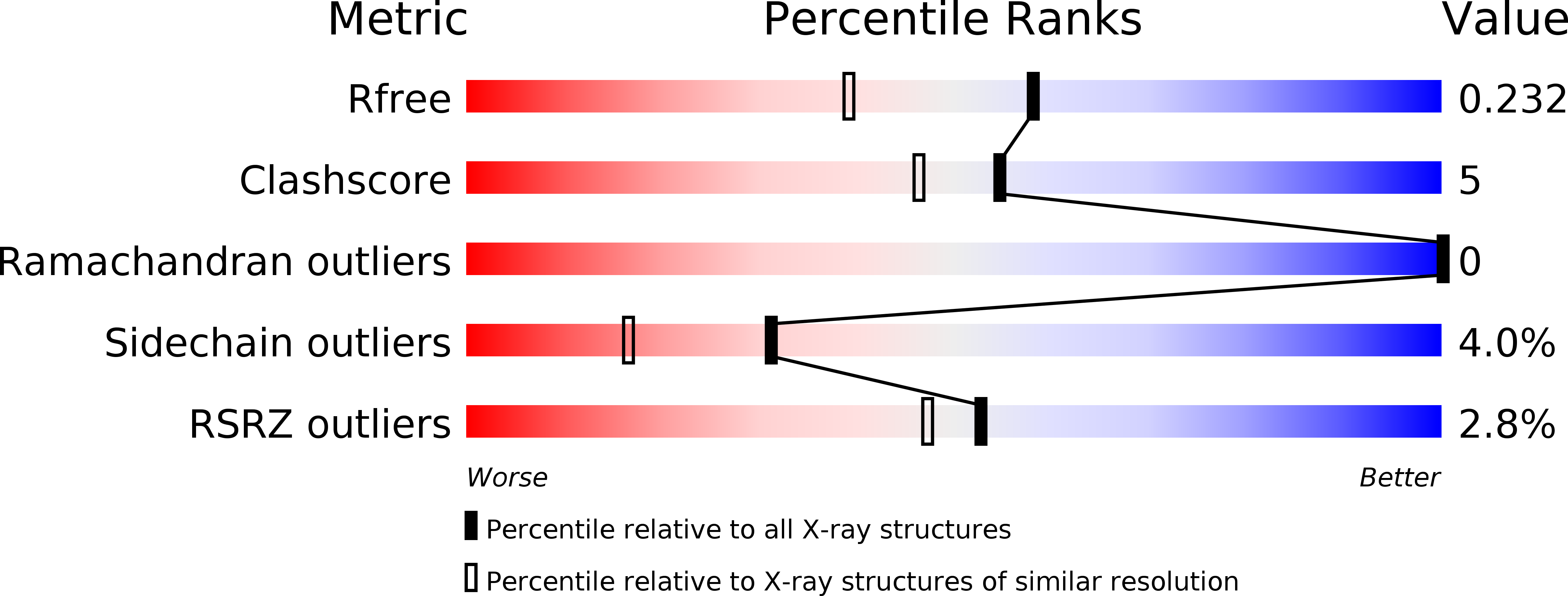
Resolution:
1.80 Å
R-Value Free:
0.23
R-Value Work:
0.18
R-Value Observed:
0.18
Space Group:
C 1 2 1
Macromolecular Entities
Structures with similar UniProt ID
Protein Blast
Polymer Type:polypeptide(L)
Molecule:3,4-dihydroxy-2-butanone 4-phosphate synthase
Gene (Uniprot):ribBA
Chain IDs:A, B
Chain Length:206
Number of Molecules:2
Biological Source:Mycobacterium tuberculosis
Primary Citation
Structural basis for pH dependent monomer-dimer transition of 3,4-dihydroxy 2-butanone-4-phosphate synthase domain from Mycobacterium tuberculosis
J.Struct.Biol. 174 374 384 (2011)
PMID: 21296160 DOI: 10.1016/j.jsb.2011.01.013

Abstact

3,4-dihydroxy 2-butanone 4-phosphate synthase (DHBPS) and GTP cyclohydrolase-II (GTPCH-II) are the two initial enzymes involved in riboflavin biosynthesis pathway, which has been shown to be essential for the pathogens. In Mycobacterium tuberculosis (Mtb), the ribA2 gene (Rv1415) encodes for the bi-functional enzyme with DHBPS and GTPCH-II domains at N- and C-termini, respectively. We have determined three crystal structures of Mtb-DHBPS domain in complex with phosphate and glycerol at pH 6.0, with sulphate at pH 4.0 and with zinc and sulphate at pH 4.0 at 1.8, 2.06 and 2.06 Å resolution, respectively. The hydrodynamic volume and enzyme activity studies revealed that the Mtb-DHBPS domain forms a functional homo-dimer between the pH 6.0 and 9.0, however, at pH 5.0 and below, it forms a stable inactive monomer in solution. Furthermore, the functional activity of Mtb-DHBPS and its dimeric state could be restored by increasing the pH between 6.0 and 9.0. The comparison of crystal structures determined at different pH revealed that the overall three-dimensional structure of Mtb-DHBPS monomer remains the same. However, the length of the α6-helix at pH 6.0 has increased from 15 to 22 Å in pH 4.0 by increasing the number of amino acids contributing to the α6-helix from 11 to 15, achieving a higher structural stability at pH 4.0. Taken together our experiments strongly suggest that the Mtb-DHBPS domain can transit between inactive monomer to active dimer depending upon its pH values, both in solution as well in crystal structure.

Legend

Protein

Chemical

Disease

Primary Citation of related structures
Feedback Form
Name
Email
Institute
Feedback