Deposition Date
2010-04-03
Release Date
2010-09-29
Last Version Date
2025-04-09
Entry Detail
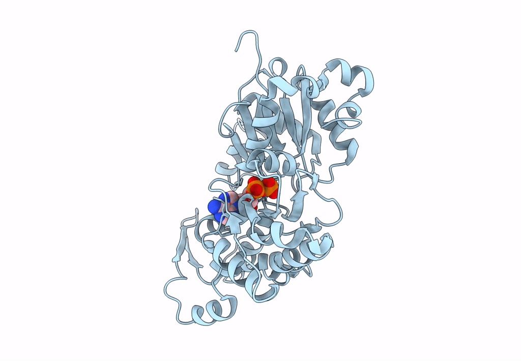
PDB ID:
3MFP
Keywords:
Title:
Atomic model of F-actin based on a 6.6 angstrom resolution cryoEM map
Biological Source:
Source Organism(s):
Oryctolagus cuniculus (Taxon ID: 9986)
Method Details:
Experimental Method:
Resolution:
6.60 Å
Aggregation State:
FILAMENT
Reconstruction Method:
HELICAL