Deposition Date
2010-03-31
Release Date
2010-04-21
Last Version Date
2023-09-06
Entry Detail
PDB ID:
3MEB
Keywords:
Title:
Structure of cytoplasmic aspartate aminotransferase from giardia lamblia
Biological Source:
Source Organism(s):
Giardia lamblia (Taxon ID: 184922)
Expression System(s):
Method Details:
Experimental Method:
Resolution:
1.90 Å
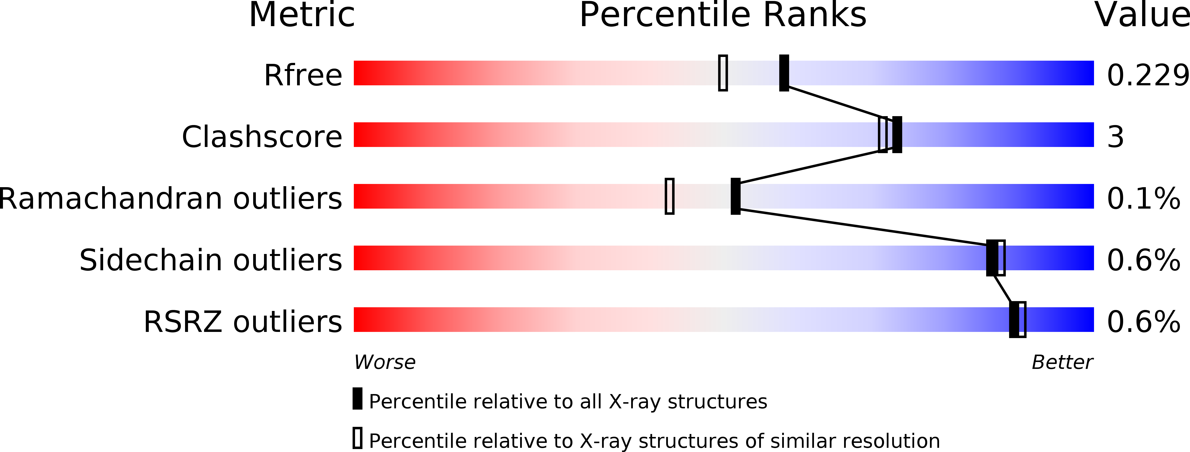
R-Value Free:
0.21
R-Value Work:
0.17
R-Value Observed:
0.17
Space Group:
P 1 21 1