Deposition Date
2010-03-26
Release Date
2010-07-28
Last Version Date
2024-11-06
Entry Detail
PDB ID:
3MC2
Keywords:
Title:
Crystal Structure of the Murine Inhibitor of Carbonic Anhydrase
Biological Source:
Source Organism(s):
Mus musculus (Taxon ID: 10090)
Expression System(s):
Method Details:
Experimental Method:
Resolution:
2.40 Å
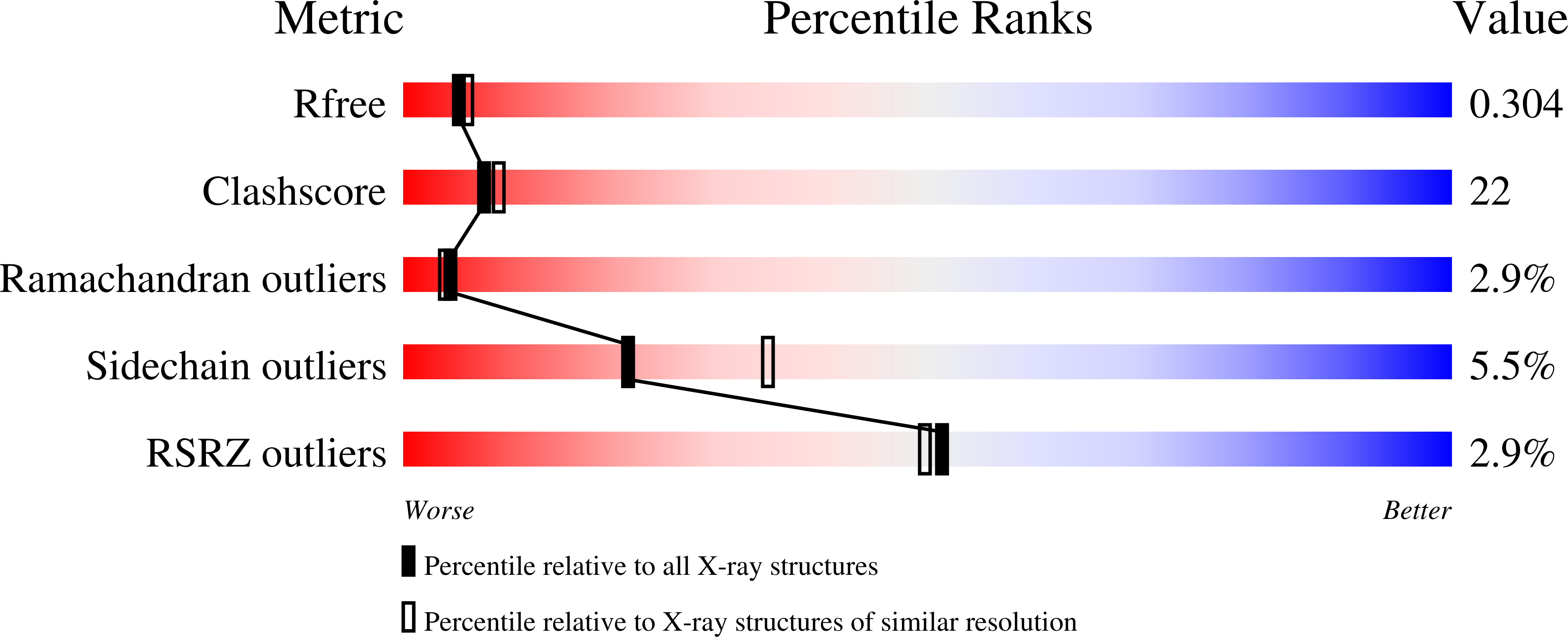
R-Value Free:
0.30
R-Value Work:
0.23
Space Group:
P 1 21 1