3MBK image
Entry Detail
PDB ID:
3MBK
Title:
The 1.35 A Structure of the Phosphatase Domain of the Suppressor of T Cell Receptor Signalling Protein in Complex with Sulphate
Biological Source:
Source Organism:
Host Organism:
PDB Version:
Deposition Date:
2010-03-25
Release Date:
2010-06-23
Method Details:
Experimental Method:
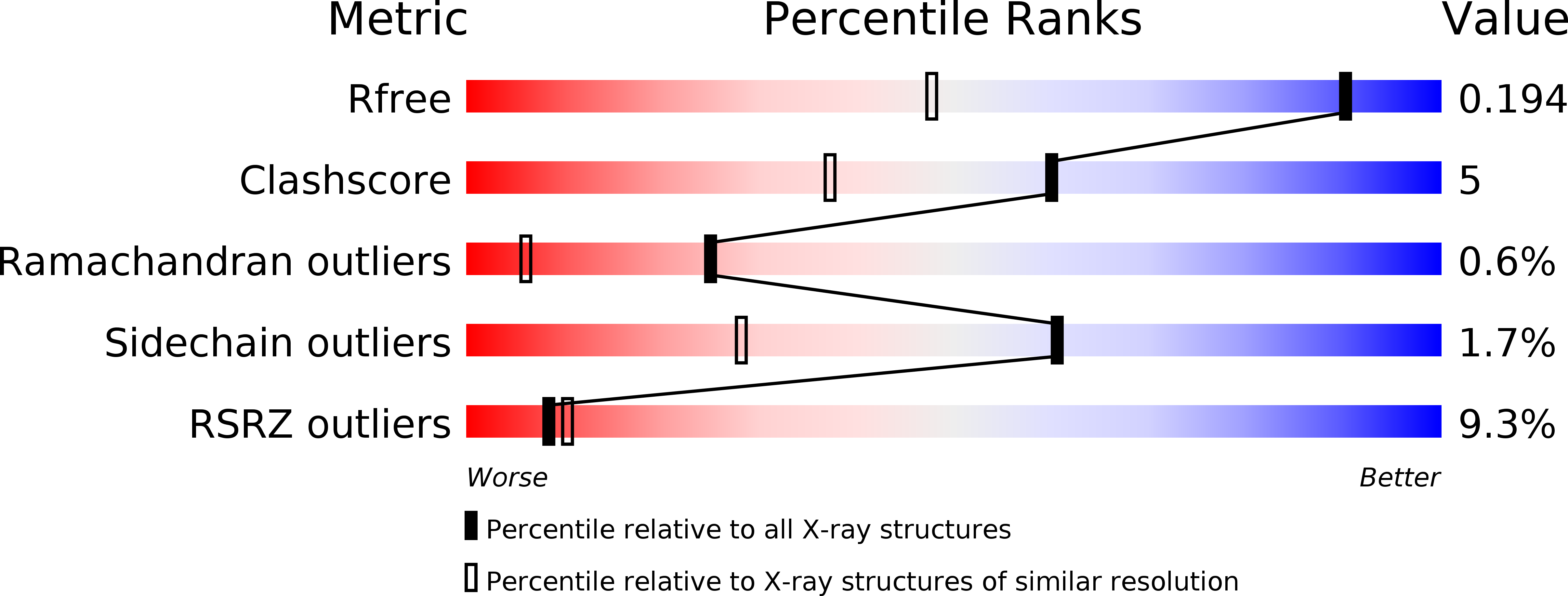
Resolution:
1.35 Å
R-Value Free:
0.19
R-Value Work:
0.16
R-Value Observed:
0.16
Space Group:
P 21 21 21
Macromolecular Entities
Polymer Type:polypeptide(L)
Description:Ubiquitin-associated and SH3 domain-containing protein B
Chain IDs:A, B
Chain Length:264
Number of Molecules:2
Biological Source:Mus musculus
Ligand Molecules
Primary Citation
The 1.35 A resolution structure of the phosphatase domain of the suppressor of T-cell receptor signaling protein in complex with sulfate.
Acta Crystallogr.,Sect.F 66 643 647 (2010)
PMID: 20516590 DOI: 10.1107/S1744309110014259

Abstact

The suppressor of T-cell signaling (Sts) proteins are multidomain proteins that negatively regulate the signaling of membrane-bound receptors, including the T-cell receptor (TCR) and the epidermal growth-factor receptor (EGFR). They contain at their C-terminus a 2H-phosphatase homology (PGM) domain that is responsible for their protein tyrosine phosphatase activity. Here, the crystal structure of the phosphatase domain of Sts-1, Sts-1(PGM), was determined at pH 4.6. The asymmetric unit contains two independent molecules and each active site is occupied by a sulfate ion. Each sulfate is located at the phosphate-binding site and makes similar interactions with the catalytic residues. The structure suggests an explanation for the lower Michaelis-Menten constants at acidic pH.

Legend

Protein

Chemical

Disease

Primary Citation of related structures