Deposition Date
2010-03-24
Release Date
2010-05-26
Last Version Date
2024-11-06
Entry Detail
PDB ID:
3MAW
Keywords:
Title:
Structure of the Newcastle disease virus F protein in the post-fusion conformation
Biological Source:
Source Organism(s):
Newcastle disease virus (Taxon ID: 11177)
Method Details:
Experimental Method:
Resolution:
3.50 Å
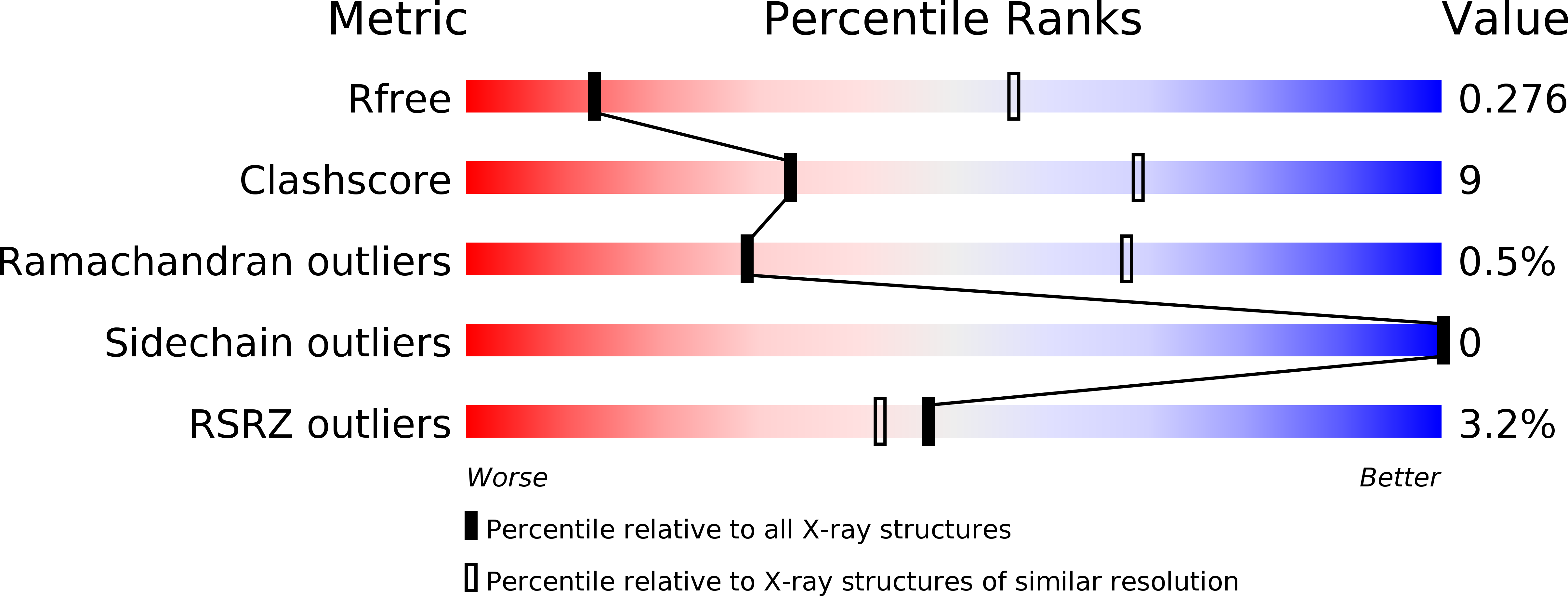
R-Value Free:
0.29
R-Value Work:
0.26
R-Value Observed:
0.26
Space Group:
H 3