3M9Z image
Deposition Date 2010-03-23
Release Date 2011-04-06
Last Version Date 2024-11-27
Entry Detail
PDB ID:
3M9Z
Title:
Crystal Structure of extracellular domain of mouse NKR-P1A
Biological Source:
Source Organism:
Mus musculus (Taxon ID: 10090)
Host Organism:
Method Details:
Experimental Method:
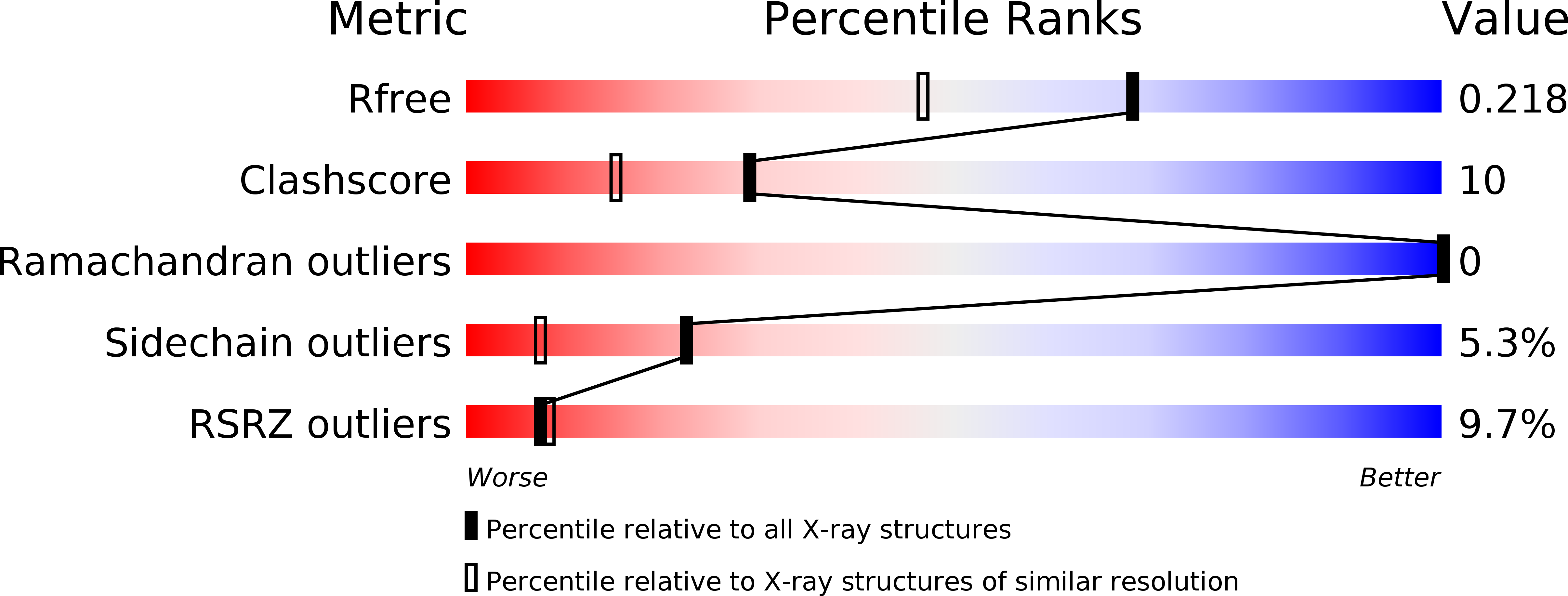
Resolution:
1.70 Å
R-Value Free:
0.23
R-Value Work:
0.19
R-Value Observed:
0.19
Space Group:
I 41 2 2
Macromolecular Entities
Polymer Type:polypeptide(L)
Molecule:Killer cell lectin-like receptor subfamily B member 1A
Gene (Uniprot):Klrb1a
Chain IDs:A
Chain Length:139
Number of Molecules:1
Biological Source:Mus musculus
Ligand Molecules
Primary Citation
Molecular architecture of mouse activating NKR-P1 receptors.
J.Struct.Biol. 175 434 441 (2011)
PMID: 21600988 DOI: 10.1016/j.jsb.2011.05.001

Abstact

Receptors belonging to NKR-P1 family and their specific Clr ligands form an alternative missing self recognition system critical in immunity against tumors and viruses, elimination of tumor cells subjected to genotoxic stress, activation of T cell dependent immune response, and hypertension. The three-dimensional structure of the extracellular domain of the mouse natural killer (NK) cell receptor mNKR-P1Aex has been determined by X-ray diffraction. The core of the C-type lectin domain (CTLD) is homologous to the other CTLD receptors whereas one quarter of the domain forms an extended loop interacting tightly with a neighboring loop in the crystal. This domain swapping mechanism results in a compact interaction interface. A second dimerization interface resembles the known arrangement of other CTLD NK receptors. A functional dimeric form of the receptor is suggested, with the loop, evolutionarily conserved within this family, proposed to participate in interactions with ligands.

Legend

Protein

Chemical

Disease

Primary Citation of related structures