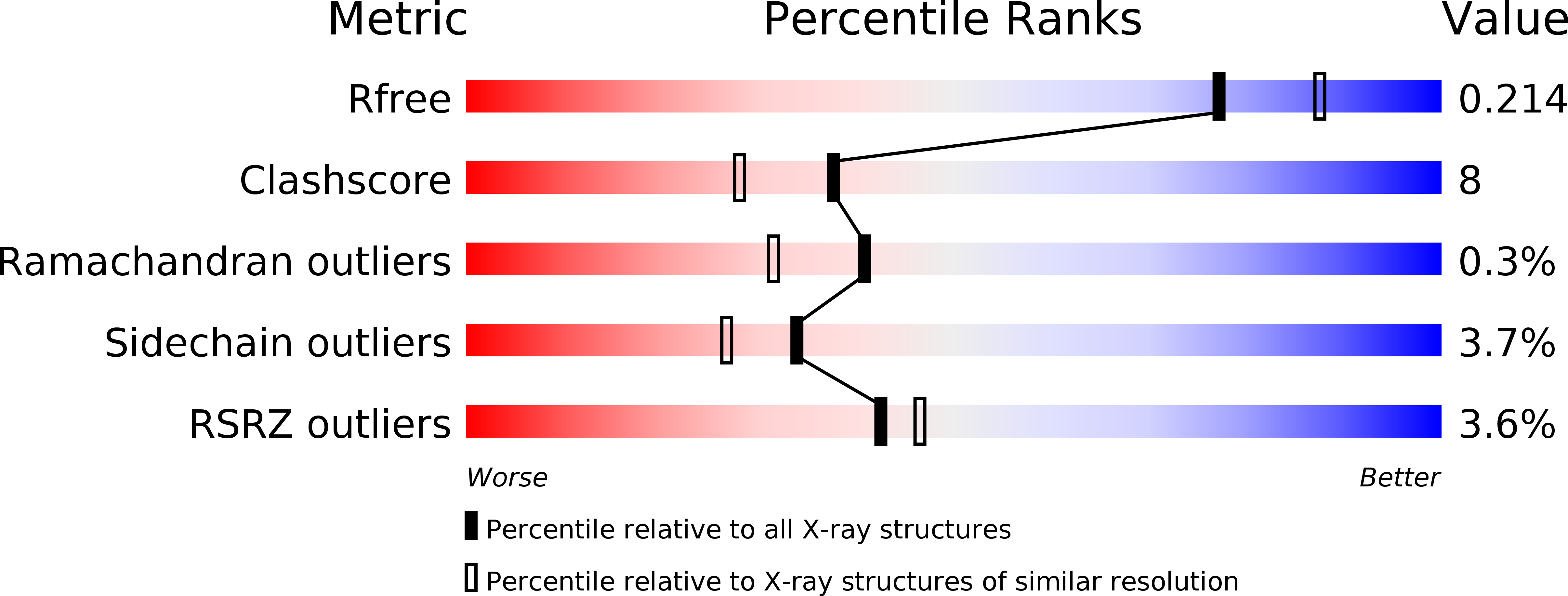
3M9V image
Deposition Date 2010-03-22
Release Date 2010-04-07
Last Version Date 2024-04-03
Entry Detail
PDB ID:
3M9V
Keywords:
Title:
X-ray Structure of a KijD3 in Complex with dTDP
Biological Source:
Source Organism:
Host Organism:
Method Details:
Experimental Method:
Resolution:
2.05 Å
R-Value Free:
0.22
R-Value Work:
0.16
R-Value Observed:
0.17
Space Group:
I 2 2 2
Macromolecular Entities
Structures with similar UniProt ID
Protein Blast
Polymer Type:polypeptide(L)
Molecule:FAD-dependent oxidoreductase
Gene (Uniprot):KijD3
Chain IDs:A
Chain Length:439
Number of Molecules:1
Biological Source:Actinomadura kijaniata
Ligand Molecules
Primary Citation
X-ray structure of kijd3, a key enzyme involved in the biosynthesis of D-kijanose.
Biochemistry 49 3517 3524 (2010)
PMID: 20334431 DOI: 10.1021/bi100318v

Abstact

D-kijanose is an unusual nitrosugar found attached to the antibiotic kijanimicin. Ten enzymes are required for its production in Actinomadura kijaniata, a soil-dwelling actinomycete. The focus of this investigation is on the protein encoded by the kijd3 gene and hereafter referred to as KijD3. On the basis of amino acid sequence analyses, KijD3 has been proposed to be an FAD-dependent oxidoreductase, which catalyzes the sixth step in d-kijanose biosynthesis by converting dTDP-3-amino-2,3,6-trideoxy-4-keto-3-methyl-d-glucose into its C-3' nitro derivative. This putative activity, however, has never been demonstrated in vivo or in vitro. Here we report the first structural study of this enzyme. For our investigation, crystals of KijD3 were grown in the presence of dTDP, and the structure was solved to 2.05-A resolution. The enzyme is a tetramer with each subunit folding into three distinct regions: a five alpha-helical bundle, an eight-stranded beta-sheet, and a second five alpha-helical bundle. The dTDP moiety is anchored to the protein via the side chains of Glu 113, Gln 254, and Arg 330. The overall fold of KijD3 places it into the well-characterized fatty acyl-CoA dehydrogenase superfamily. There is a decided cleft in each subunit with the appropriate dimensions to accommodate a dTDP-linked sugar. Strikingly, the loop defined by Phe 383 to Ala 388, which projects into the active site, contains two adjacent cis-peptide bonds, Pro 386 and Tyr 387. Activity assays demonstrate that KijD3 requires FAD for activity and that it produces a hydroxylamino product. The molecular architecture of KijD3 described in this report serves as a paradigm for a new family of enzymes that function on dTDP-linked sugar substrates.

Legend

Protein

Chemical

Disease

Primary Citation of related structures