3M9E image
Deposition Date 2010-03-22
Release Date 2010-08-18
Last Version Date 2023-09-06
Entry Detail
PDB ID:
3M9E
Title:
Thyroid hormone beta DNA binding domain homodimer with inverted palindrome TRE
Biological Source:
Source Organism:
Host Organism:
Method Details:
Experimental Method:
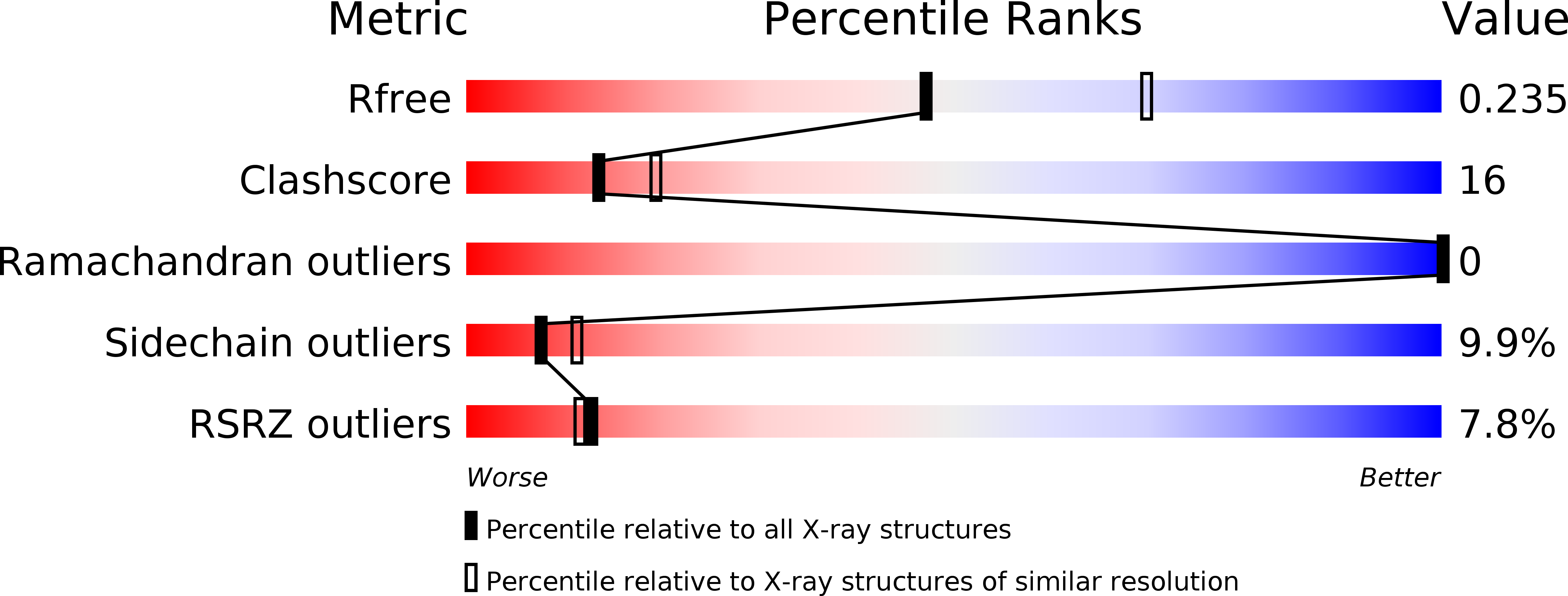
Resolution:
2.41 Å
R-Value Free:
0.23
R-Value Work:
0.17
R-Value Observed:
0.18
Space Group:
P 1 21 1
Macromolecular Entities
Structures with similar UniProt ID
Protein Blast
Polymer Type:polypeptide(L)
Molecule:Thyroid hormone receptor beta
Gene (Uniprot):Thrb
Chain IDs:A, B, E, F
Chain Length:105
Number of Molecules:4
Biological Source:Rattus norvegicus
Polymer Type:polydeoxyribonucleotide
Molecule:DNA (5'-D(*AP*TP*TP*GP*AP*CP*CP*TP*CP*AP*GP*CP*TP*GP*AP*GP*GP*TP*CP*AP*AP*T)-3')
Chain IDs:C, D, G, H
Chain Length:22
Number of Molecules:4
Biological Source:
Ligand Molecules
Primary Citation
Structure of a thyroid hormone receptor DNA-binding domain homodimer bound to an inverted palindrome DNA response element.
Mol.Endocrinol. 24 1650 1664 (2010)
PMID: 20610536 DOI: 10.1210/me.2010-0129

Abstact

Thyroid hormone receptor (TR), as a member of the nuclear hormone receptor family, can recognize and bind different classes of DNA response element targets as either a monomer, a homooligomer, or a heterooligomer. We report here the first crystal structure of a homodimer TR DNA-binding domain (DBD) in complex with an inverted repeat class of thyroid response element (TRE). The structure shows a nearly symmetric structure of the TR DBD assembled on the F2 TRE where the base recognition contacts in the homodimer DNA complex are conserved relative to the previously published structure of a TR-9-cis-retinoic acid receptor heterodimer DNA complex. The new structure also reveals that the T-box region of the DBD can function as a structural hinge that enables a large degree of flexibility in the position of the C-terminal extension helix that connects the DBD to the ligand-binding domain. Although the isolated TR DBDs exist as monomers in solution, we have measured highly cooperative binding of the two TR DBD subunits onto the inverted repeat DNA sequence. This suggests that elements of the DBD can influence the specific TR oligomerization at target genes, and it is not just interactions between the ligand-binding domains that are responsible for TR oligomerization at target genes. Mutational analysis shows that intersubunit contacts at the DBD C terminus account for some, but not all, of the cooperative homodimer TR binding to the inverted repeat class TRE.

Legend

Protein

Chemical

Disease

Primary Citation of related structures
Feedback Form
Name
Email
Institute
Feedback