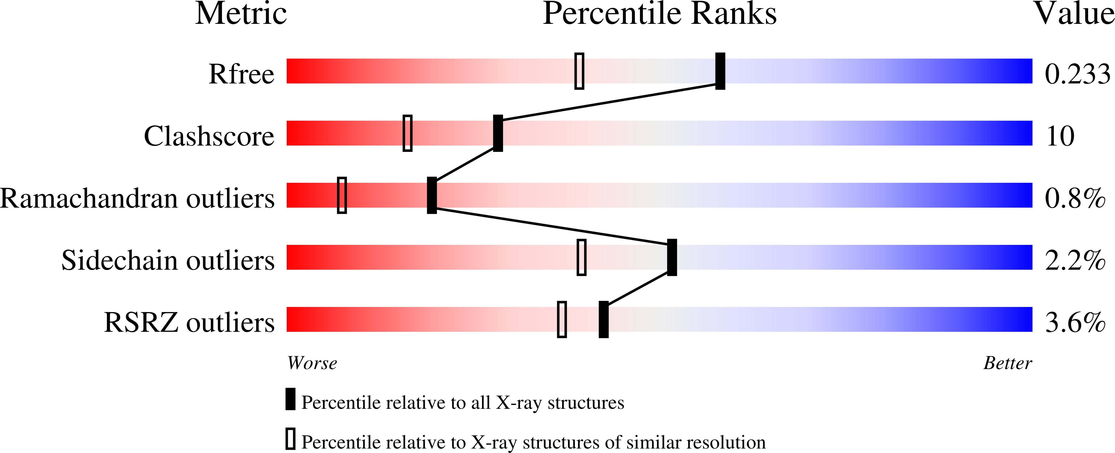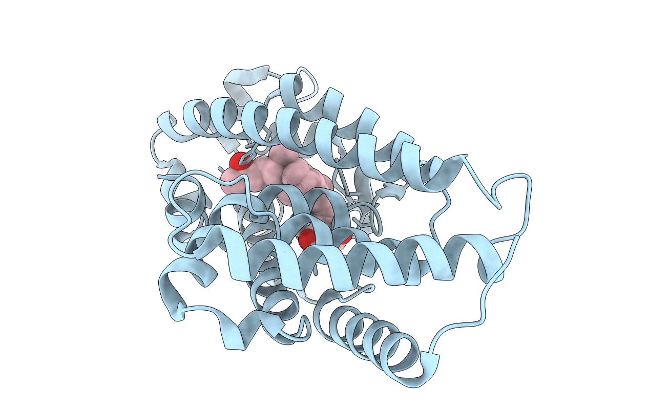
3M7R image
Entry Detail
PDB ID:
3M7R
Keywords:
Title:
Crystal structure of VDR H305Q mutant
Biological Source:
Source Organism:
Host Organism:
PDB Version:
Deposition Date:
2010-03-17
Release Date:
2010-05-05
Method Details:
Experimental Method:
Resolution:
1.80 Å
R-Value Free:
0.24
R-Value Work:
0.21
R-Value Observed:
0.24
Space Group:
P 21 21 21
Macromolecular Entities
Polymer Type:polypeptide(L)
Description:Vitamin D3 receptor
Mutations:H305Q
Chain IDs:A
Chain Length:253
Number of Molecules:1
Biological Source:Homo sapiens
Ligand Molecules
Primary Citation
Crystal structure of hereditary vitamin D-resistant rickets--associated mutant H305Q of vitamin D nuclear receptor bound to its natural ligand
J.Steroid Biochem.Mol.Biol. 121 84 87 (2010)
PMID: 20403435 DOI: 10.1016/j.jsbmb.2010.04.008

Abstact

In the nuclear receptor of vitamin D (VDR) histidine 305 participates to the anchoring of the ligand. The VDR H305Q mutation was identified in a patient who exhibited the hereditary vitamin D-resistant rickets (HVDRR). We report the crystal structure of human VDR H305Q-ligand binding domain bound to 1alpha,25(OH)2D3 solved at 1.8A resolution. The protein adopts the active conformation of the wild-type liganded VDR. A local conformational flexibility at the mutation site weakens the hydrogen bond between the 25-OH with Gln305, thus explaining the lower affinity of the mutant proteins for calcitriol. The structure provides the basis for a rational approach to the design of more potent ligands for the treatment of HVDRR.

Legend

Protein

Chemical

Disease

Primary Citation of related structures