Deposition Date
2010-03-17
Release Date
2011-03-16
Last Version Date
2024-11-27
Entry Detail
PDB ID:
3M7Q
Keywords:
Title:
Crystal structure of recombinant Kunitz Type serine protease Inhibitor-1 from the Caribbean sea anemone stichodactyla helianthus in complex with bovine pancreatic trypsin
Biological Source:
Source Organism(s):
Stichodactyla helianthus (Taxon ID: 6123)
Bos taurus (Taxon ID: 9913)
Bos taurus (Taxon ID: 9913)
Expression System(s):
Method Details:
Experimental Method:
Resolution:
1.70 Å
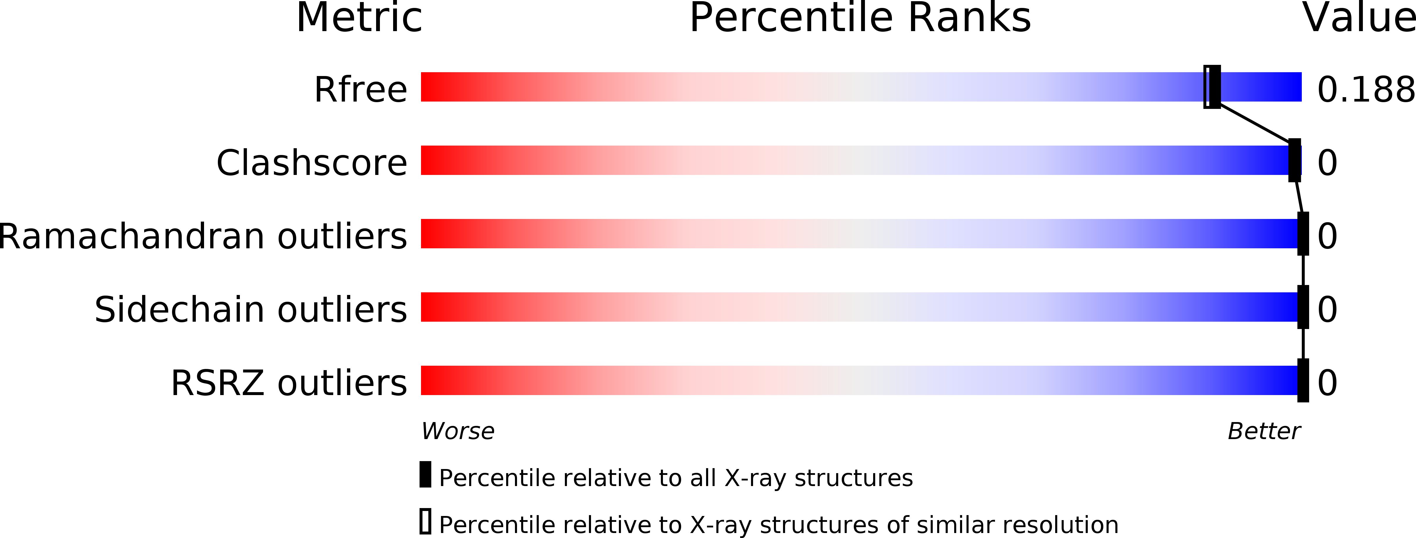
R-Value Free:
0.18
R-Value Work:
0.15
R-Value Observed:
0.15
Space Group:
P 21 21 21