3M7O image
Entry Detail
PDB ID:
3M7O
Keywords:
Title:
Crystal structure of mouse MD-1 in complex with phosphatidylcholine
Biological Source:
Source Organism:
Host Organism:
PDB Version:
Deposition Date:
2010-03-17
Release Date:
2010-06-09
Method Details:
Experimental Method:
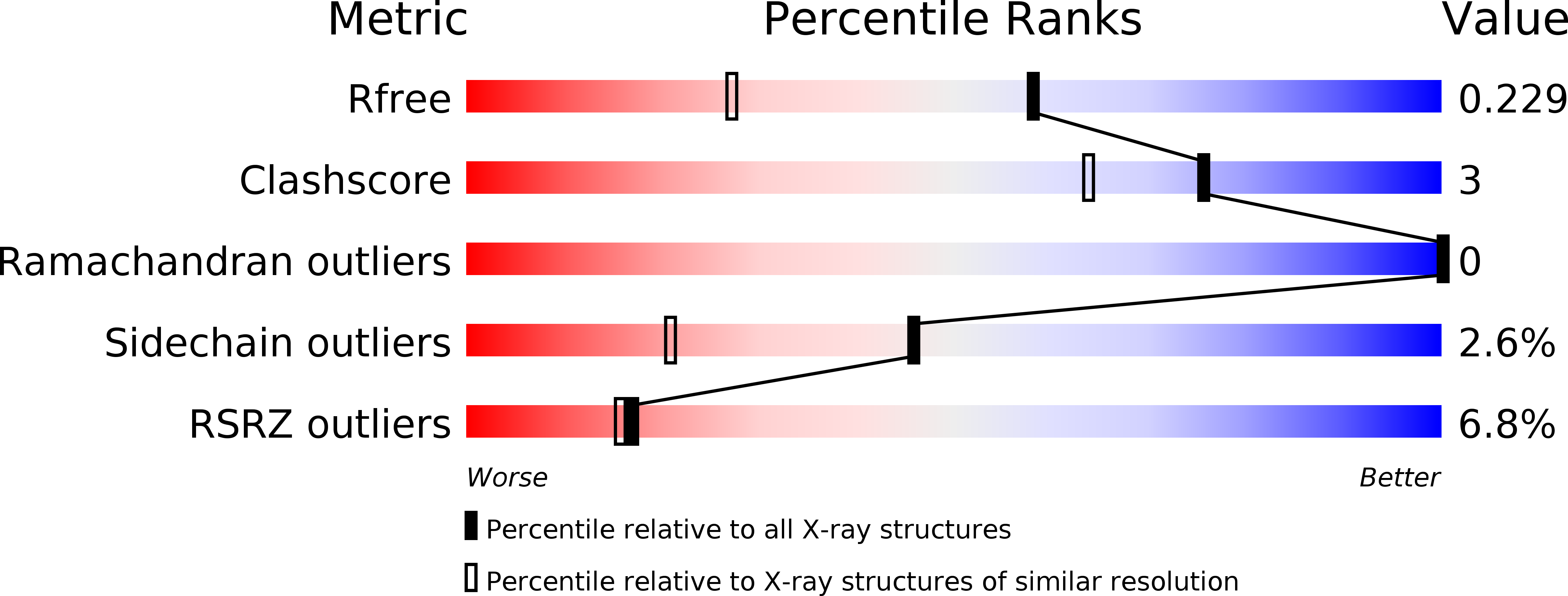
Resolution:
1.65 Å
R-Value Free:
0.22
R-Value Work:
0.19
R-Value Observed:
0.19
Space Group:
P 43
Macromolecular Entities
Polymer Type:polypeptide(L)
Description:Lymphocyte antigen 86
Chain IDs:A, B, C, D
Chain Length:162
Number of Molecules:4
Biological Source:Mus musculus
Modified Residue
Compound ID Chain ID Parent Comp ID Details 2D Image
ASN A ASN GLYCOSYLATION SITE
Primary Citation
Crystal structure of mouse MD-1 with endogenous phospholipid bound in its cavity
J.Mol.Biol. 400 838 846 (2010)
PMID: 20595044 DOI: 10.1016/j.jmb.2010.05.063

Abstact

MD-1 is a glycoprotein that associates with a B-cell-specific RP105 protein and has a low sequence identity of 16% to MD-2 that associates with Toll-like receptor 4 and recognizes endotoxic lipopolysaccharide. MD-1 and RP105 are supposed to mediate lipopolysaccharide recognition; however, little is known about their structures and functions. Here, the crystal structure of mouse MD-1 is determined at 1.65 A resolution. MD-1 has a hydrophobic cavity sandwiched by two beta-sheets as is MD-2. The cavity is 25 A long, 5 A wide, and 10 A deep: longer, narrower, and shallower than that of MD-2. No charged residues are located on the cavity entrance. MD-1 is primarily monomeric in solution but shows a dimeric assembly in the crystal lattices, with their cavity entrances facing each other. In the cavity, electron densities attributable to phosphatidylcholine are located. Together with the binding assay with tetra-acylated lipid IVa, MD-1 is shown to be a lipid-binding coreceptor.

Legend

Protein

Chemical

Disease

Primary Citation of related structures