3M7D image
Deposition Date 2010-03-16
Release Date 2010-08-04
Last Version Date 2024-11-27
Entry Detail
PDB ID:
3M7D
Keywords:
Title:
Crystal structure of an N-terminal 44 kDA fragment of topoisomerase V in the presence of dioxane
Biological Source:
Source Organism:
Host Organism:
Method Details:
Experimental Method:
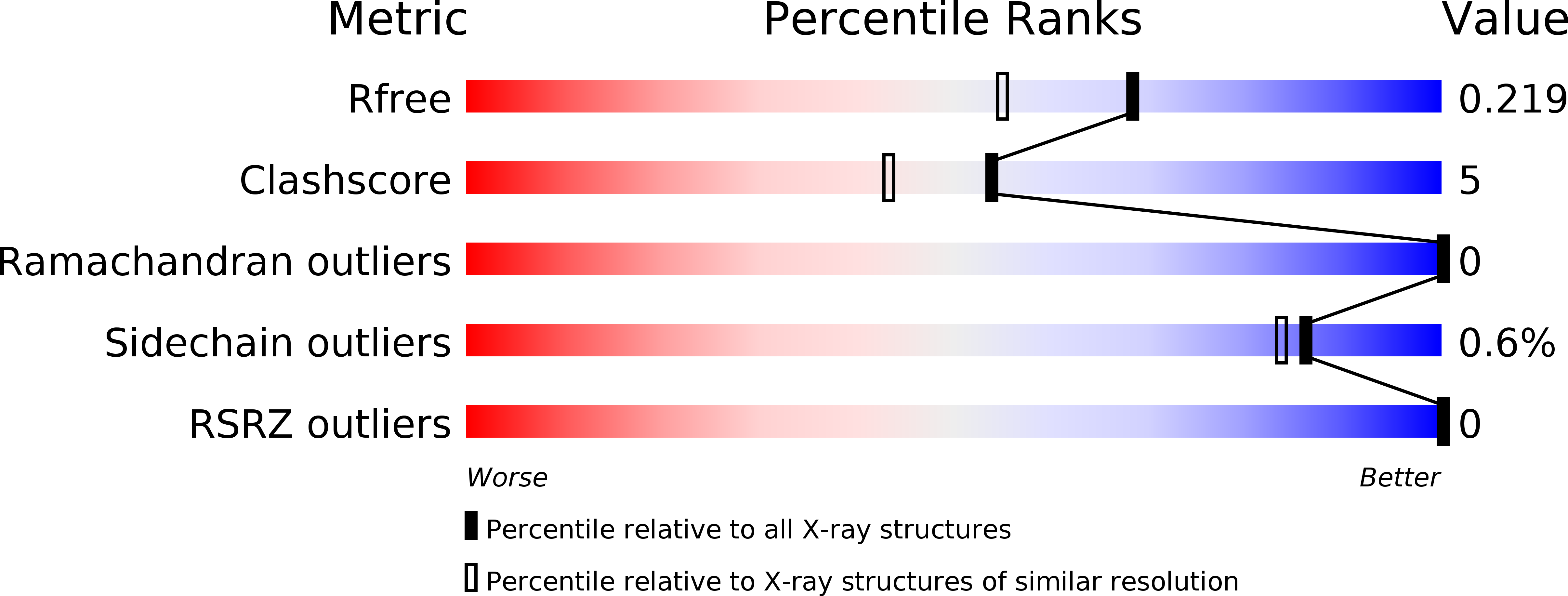
Resolution:
1.82 Å
R-Value Free:
0.22
R-Value Work:
0.17
R-Value Observed:
0.17
Space Group:
C 1 2 1
Macromolecular Entities
Polymer Type:polypeptide(L)
Molecule:Topoisomerase V
Gene (Uniprot):top5
Chain IDs:A
Chain Length:380
Number of Molecules:1
Biological Source:Methanopyrus kandleri
Primary Citation
Structures of minimal catalytic fragments of topoisomerase v reveals conformational changes relevant for DNA binding.
Structure 18 829 838 (2010)
PMID: 20637419 DOI: 10.1016/j.str.2010.03.006

Abstact

Topoisomerase V is an archaeal type I topoisomerase that is unique among topoisomerases due to presence of both topoisomerase and DNA repair activities in the same protein. It is organized as an N-terminal topoisomerase domain followed by 24 tandem helix-hairpin-helix (HhH) motifs. Structural studies have shown that the active site is buried by the (HhH) motifs. Here we show that the N-terminal domain can relax DNA in the absence of any HhH motifs and that the HhH motifs are required for stable protein-DNA complex formation. Crystal structures of various topoisomerase V fragments show changes in the relative orientation of the domains mediated by a long bent linker helix, and these movements are essential for the DNA to enter the active site. Phosphate ions bound to the protein near the active site helped model DNA in the topoisomerase domain and show how topoisomerase V may interact with DNA.

Legend

Protein

Chemical

Disease

Primary Citation of related structures