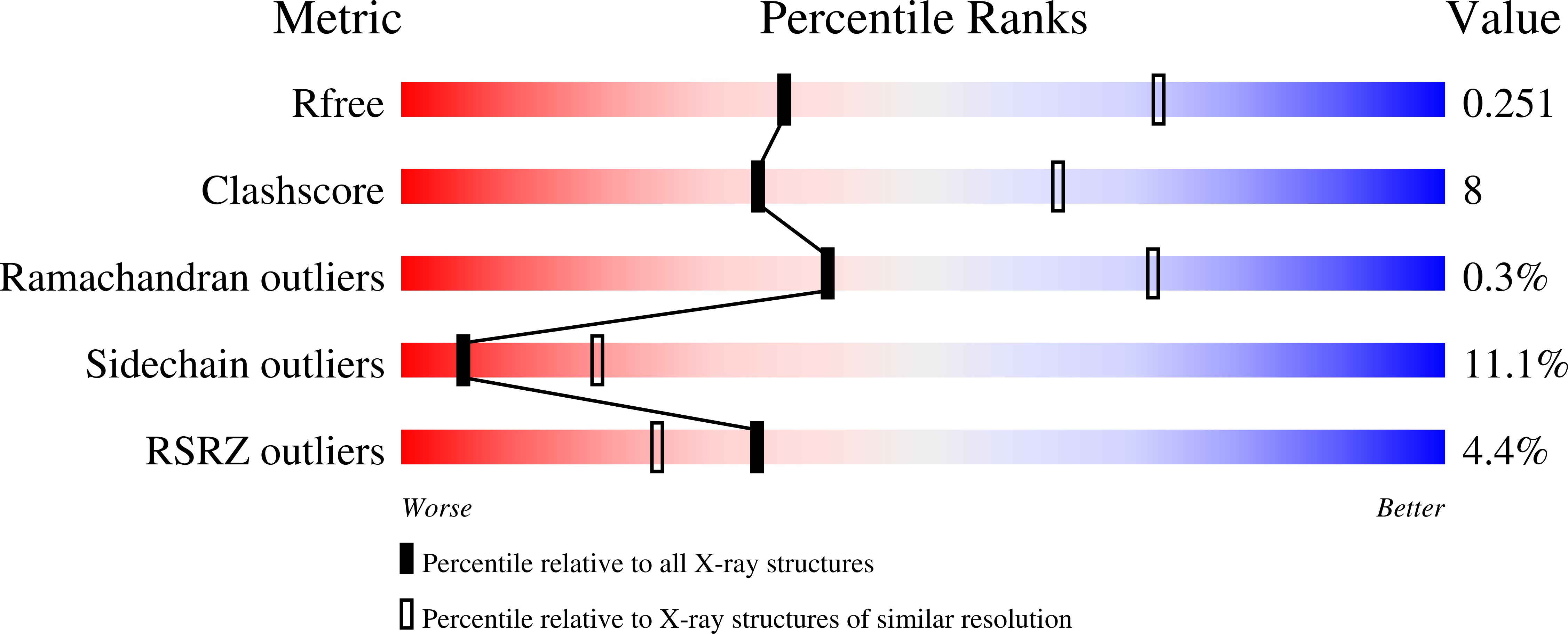
3M6S image
Deposition Date 2010-03-16
Release Date 2010-03-31
Last Version Date 2024-11-27
Entry Detail
PDB ID:
3M6S
Keywords:
Title:
Crystal structure of H1N1pdm Hemagglutinin
Biological Source:
Source Organism:
Host Organism:
Method Details:
Experimental Method:
Resolution:
2.80 Å
R-Value Free:
0.25
R-Value Work:
0.22
R-Value Observed:
0.23
Space Group:
P 1
Macromolecular Entities
Structures with similar UniProt ID
Protein Blast
Polymer Type:polypeptide(L)
Molecule:Hemagglutinin
Gene (Uniprot):HA
Chain IDs:A, C, E, G, I, K
Chain Length:331
Number of Molecules:6
Biological Source:Influenza A virus
Structures with similar UniProt ID
Protein Blast
Polymer Type:polypeptide(L)
Molecule:Hemagglutinin
Gene (Uniprot):HA
Chain IDs:B, D, F, H, J, L
Chain Length:181
Number of Molecules:6
Biological Source:Influenza A virus
Modified Residue
Compound ID Chain ID Parent Comp ID Details 2D Image
ASN E ASN GLYCOSYLATION SITE
Ligand Molecules
Primary Citation
Structure and Receptor binding properties of a pandemic H1N1 virus hemagglutinin
PLoS Curr ? RRN1152 RRN1152 (2010)
PMID: 20352039

Abstact

The 3D-structure of the major surface viral antigen from the recent H1N1 pandemic influenza virus (A/Darwin/2001/2009) was determined to 2.8 Å resolution. The structure was used to analyze changes in the HA that have emerged during the first 11 months of the pandemic and have raised public health concerns. Receptor binding properties of this protein reveals a strict preference for human-type receptors.

Legend

Protein

Chemical

Disease

Primary Citation of related structures