Deposition Date
2010-03-15
Release Date
2010-05-05
Last Version Date
2024-02-21
Entry Detail
PDB ID:
3M66
Keywords:
Title:
Crystal structure of human Mitochondrial Transcription Termination Factor 3
Biological Source:
Source Organism(s):
Homo sapiens (Taxon ID: 9606)
Expression System(s):
Method Details:
Experimental Method:
Resolution:
1.60 Å
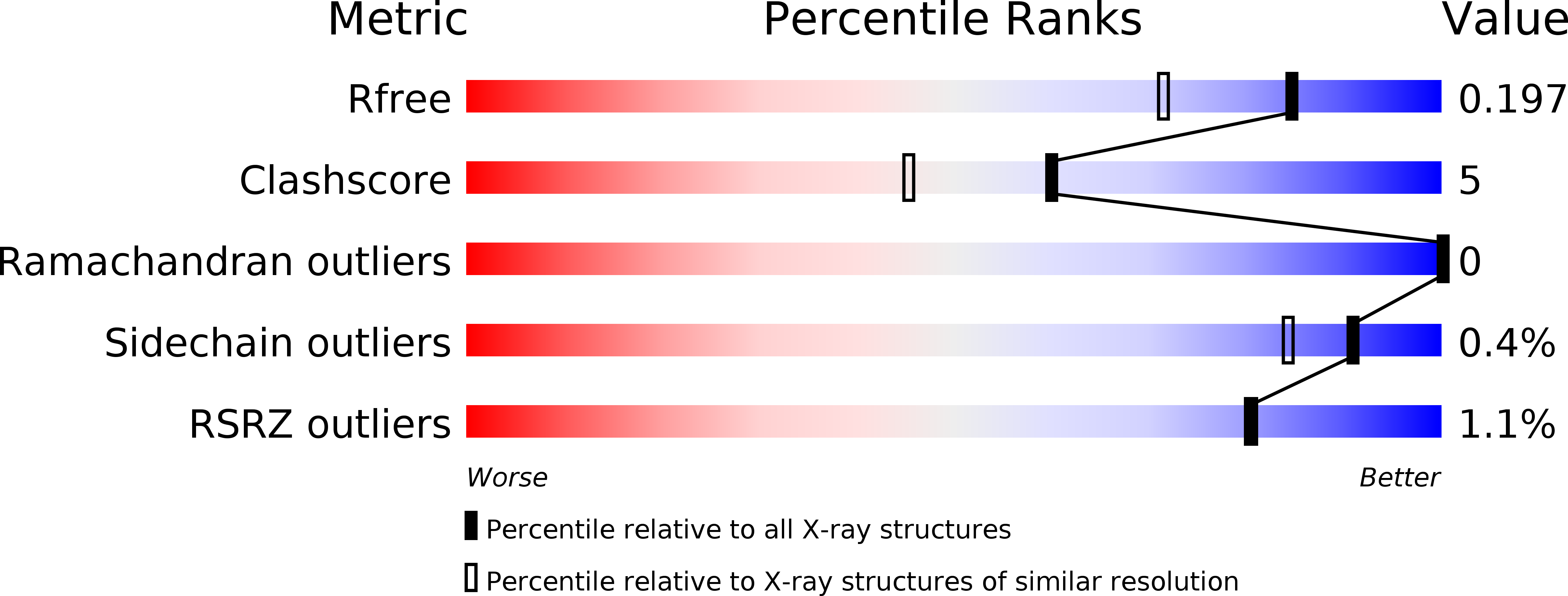
R-Value Free:
0.19
R-Value Work:
0.17
R-Value Observed:
0.17
Space Group:
C 1 2 1