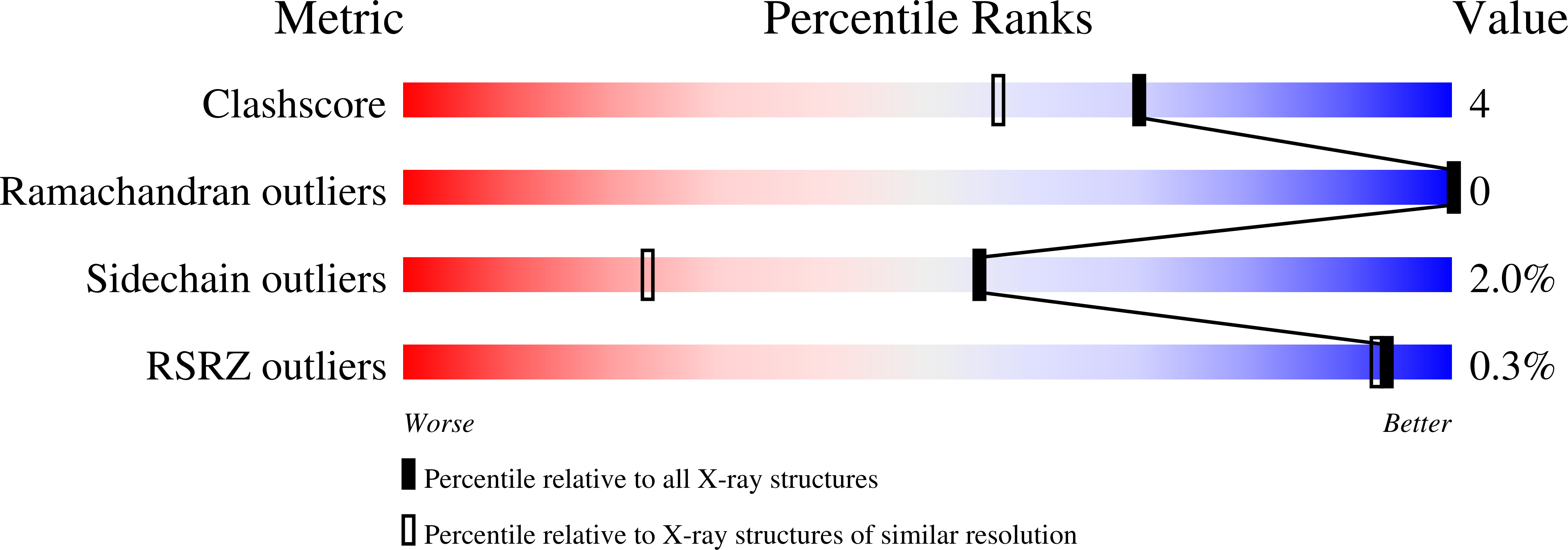
3M2G image
Deposition Date 2010-03-07
Release Date 2010-05-12
Last Version Date 2024-02-21
Entry Detail
PDB ID:
3M2G
Keywords:
Title:
Crystallographic and Single Crystal Spectral Analysis of the Peroxidase Ferryl Intermediate
Biological Source:
Source Organism:
Host Organism:
Method Details:
Experimental Method:
Resolution:
1.40 Å
R-Value Work:
0.11
Space Group:
P 21 21 21
Macromolecular Entities
Polymer Type:polypeptide(L)
Molecule:Cytochrome c peroxidase, mitochondrial
Gene (Uniprot):CCP1
Mutations:N184R
Chain IDs:A
Chain Length:291
Number of Molecules:1
Biological Source:Saccharomyces cerevisiae
Primary Citation
Crystallographic and single-crystal spectral analysis of the peroxidase ferryl intermediate.
Biochemistry 49 2984 2986 (2010)
PMID: 20230048 DOI: 10.1021/bi100238r

Abstact

The ferryl [Fe(IV)O] intermediate is important in many heme enzymes, and thus, the precise nature of the Fe(IV)-O bond is critical in understanding enzymatic mechanisms. The 1.40 A crystal structure of cytochrome c peroxidase Compound I has been determined as a function of X-ray dose while the visible spectrum was being monitored. The Fe-O bond increases in length from 1.73 A in the low-X-ray dose structure to 1.90 A in the high-dose structure. The low-dose structure correlates well with an Fe(IV) horizontal lineO bond, while we postulate that the high-dose structure is the cryo-trapped Fe(III)-OH species previously thought to be an Fe(IV)-OH species.

Legend

Protein

Chemical

Disease

Primary Citation of related structures