3M1S image
Deposition Date 2010-03-05
Release Date 2010-12-22
Last Version Date 2023-09-06
Entry Detail
PDB ID:
3M1S
Title:
Structure of Ruthenium Half-Sandwich Complex Bound to Glycogen Synthase Kinase 3
Biological Source:
Source Organism:
Homo sapiens (Taxon ID: 9606)
Host Organism:
Method Details:
Experimental Method:
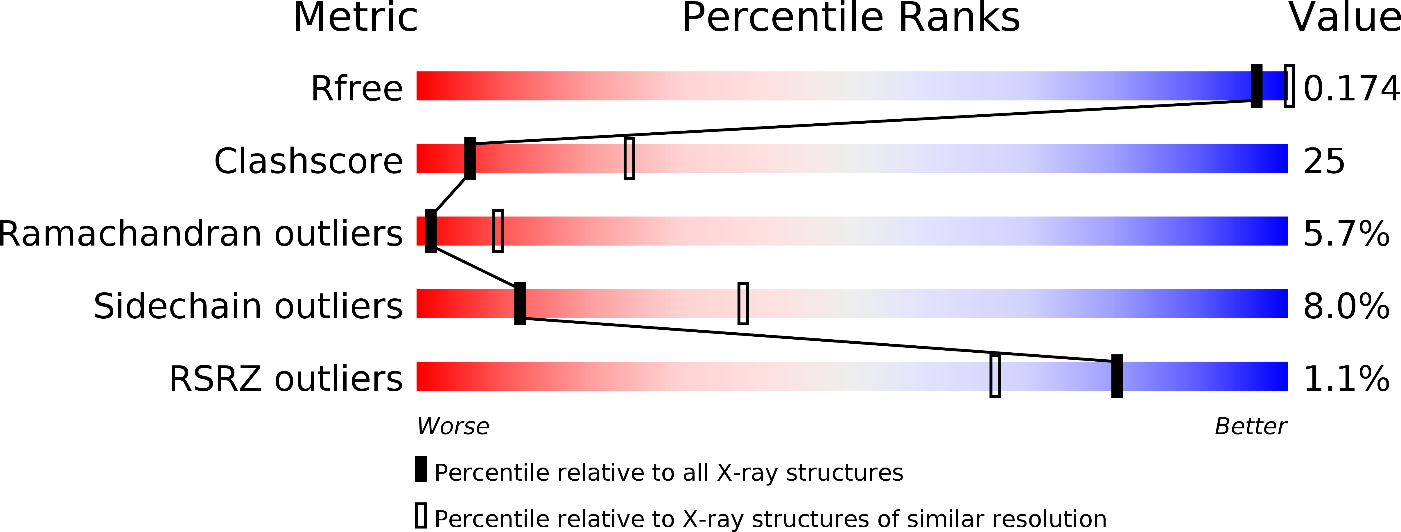
Resolution:
3.13 Å
R-Value Free:
0.22
R-Value Work:
0.16
R-Value Observed:
0.17
Space Group:
P 21 21 21
Macromolecular Entities
Structures with similar UniProt ID
Protein Blast
Polymer Type:polypeptide(L)
Molecule:Glycogen synthase kinase-3 beta
Gene (Uniprot):GSK3B
Chain IDs:A, B
Chain Length:420
Number of Molecules:2
Biological Source:Homo sapiens
Ligand Molecules
Primary Citation
Structure of anticancer ruthenium half-sandwich complex bound to glycogen synthase kinase 3beta
J.Biol.Inorg.Chem. 16 45 50 (2011)
PMID: 20821241 DOI: 10.1007/s00775-010-0699-x

Abstact

The 3.15-Å-resolution crystal structure of the R enantiomer of the highly bioactive and antiproliferative half-sandwich ruthenium complex DW12 bound to the ATP binding site of glycogen synthase kinase 3β (GSK-3β) is reported and the binding is compared with the GSK-3β binding of staurosporine and other organic inhibitors. The structure reveals a close packing of the organometallic inhibitor in the ATP binding site of GSK-3β via an induced-fit mechanism. The molecular structure of (R)-DW12 with the CO ligand oriented perpendicular to the pyridocarbazole heterocycle allows the complex to stretch the whole distance sandwiched between the faces of the N- and C-terminal lobes and to interact tightly with the flexible glycine-rich loop, which is uncommon for the interaction of GSK-3β with organic inhibitors.

Legend

Protein

Chemical

Disease

Primary Citation of related structures