Deposition Date
2010-03-04
Release Date
2010-09-15
Last Version Date
2023-09-06
Entry Detail
PDB ID:
3M1F
Keywords:
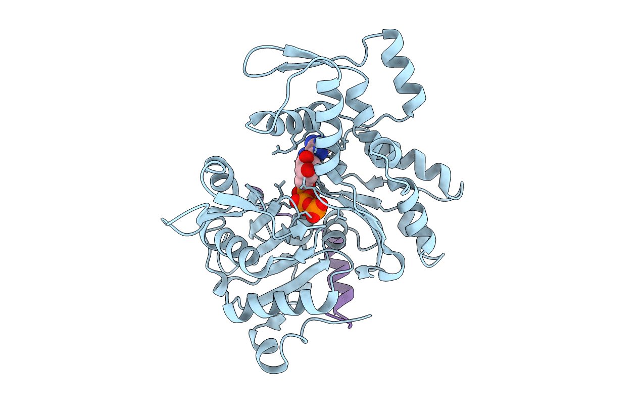
Title:
Crosslinked complex of actin with first W domain of Vibrio parahaemolyticus VopL
Biological Source:
Source Organism(s):
Vibrio parahaemolyticus (Taxon ID: 670)
Oryctolagus cuniculus (Taxon ID: 9986)
Oryctolagus cuniculus (Taxon ID: 9986)
Method Details:
Experimental Method:
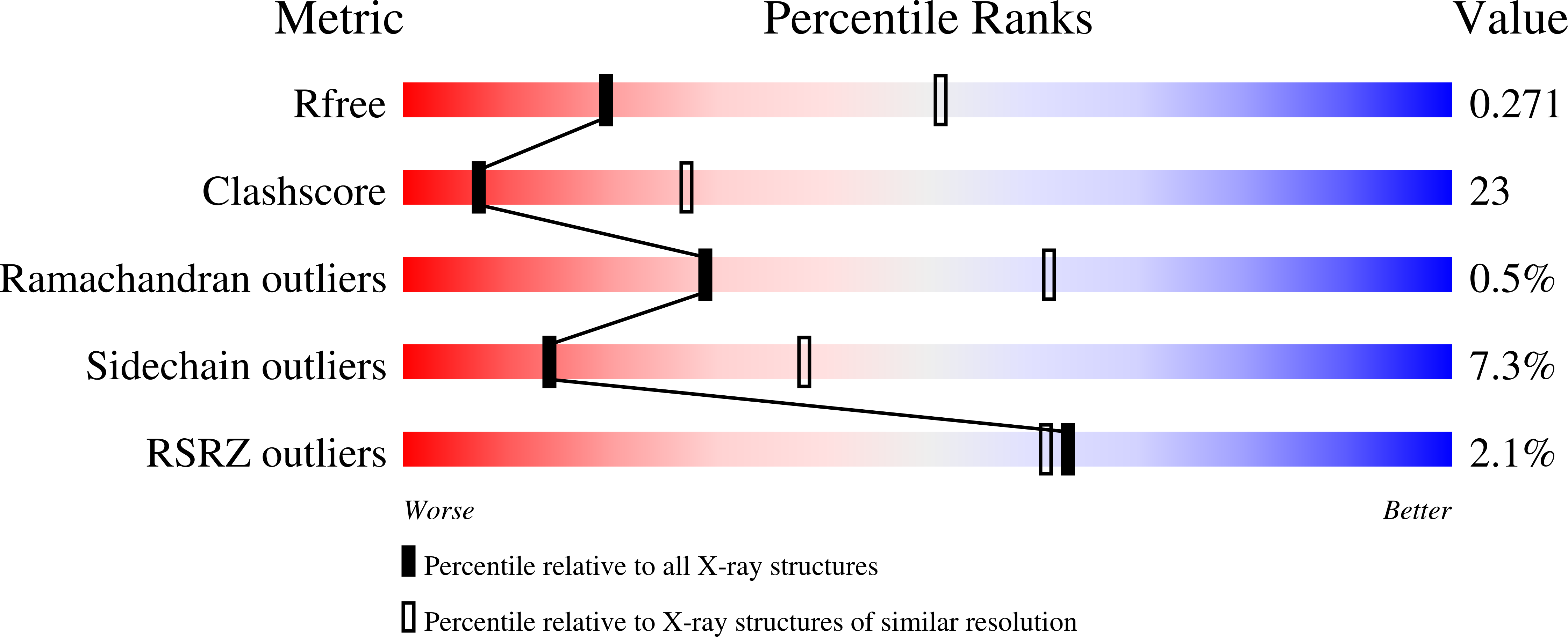
Resolution:
2.89 Å
R-Value Free:
0.26
R-Value Work:
0.21
R-Value Observed:
0.21
Space Group:
P 21 21 21