Deposition Date
2010-03-04
Release Date
2010-07-07
Last Version Date
2024-11-06
Entry Detail
PDB ID:
3M1C
Keywords:
Title:
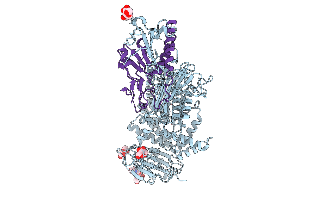
Crystal structure of the conserved herpesvirus fusion regulator complex gH-gL
Biological Source:
Source Organism(s):
Human herpesvirus 2 (Taxon ID: 10315)
Expression System(s):
Method Details:
Experimental Method:
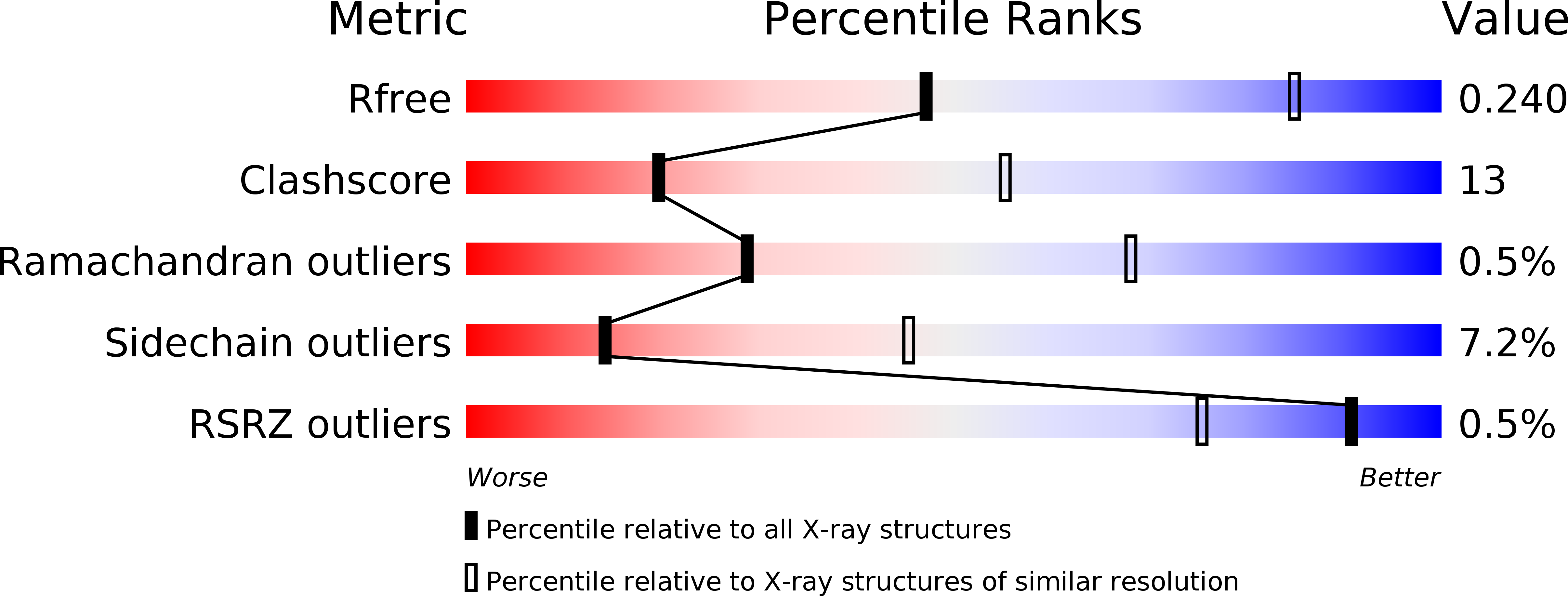
Resolution:
3.00 Å
R-Value Free:
0.24
R-Value Work:
0.17
R-Value Observed:
0.17
Space Group:
P 41 21 2