
Deposition Date
2010-02-28
Release Date
2010-03-31
Last Version Date
2024-03-20
Entry Detail
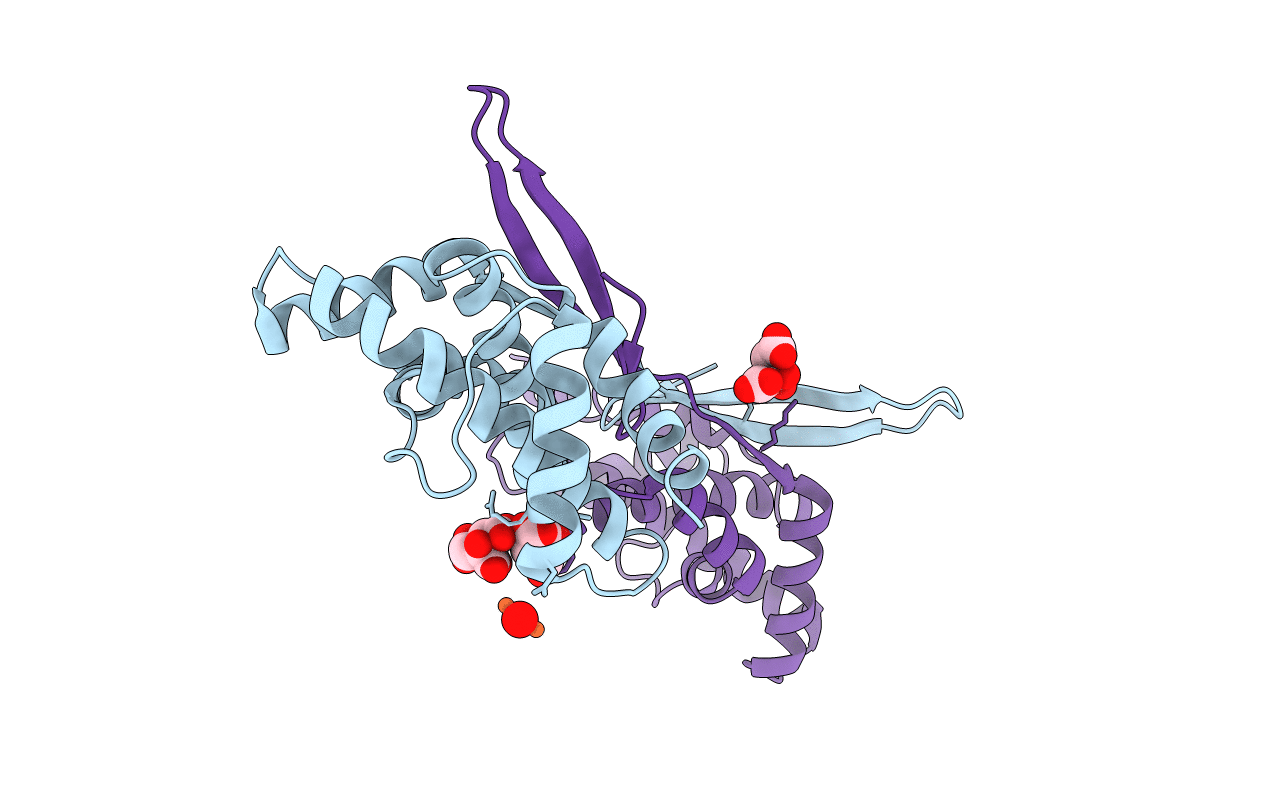
PDB ID:
3LYQ
Keywords:
Title:
Crystal structure of IpgB2 from Shigella flexneri
Biological Source:
Source Organism:
Shigella flexneri (Taxon ID: 623)
Host Organism:
Method Details:
Experimental Method:
Resolution:
2.30 Å
R-Value Free:
0.29
R-Value Work:
0.22
R-Value Observed:
0.23
Space Group:
P 4 21 2
