3LY8 image
Deposition Date 2010-02-26
Release Date 2011-02-09
Last Version Date 2024-10-30
Entry Detail
PDB ID:
3LY8
Title:
Crystal structure of mutant D471E of the periplasmic domain of CadC
Biological Source:
Source Organism:
Host Organism:
Method Details:
Experimental Method:
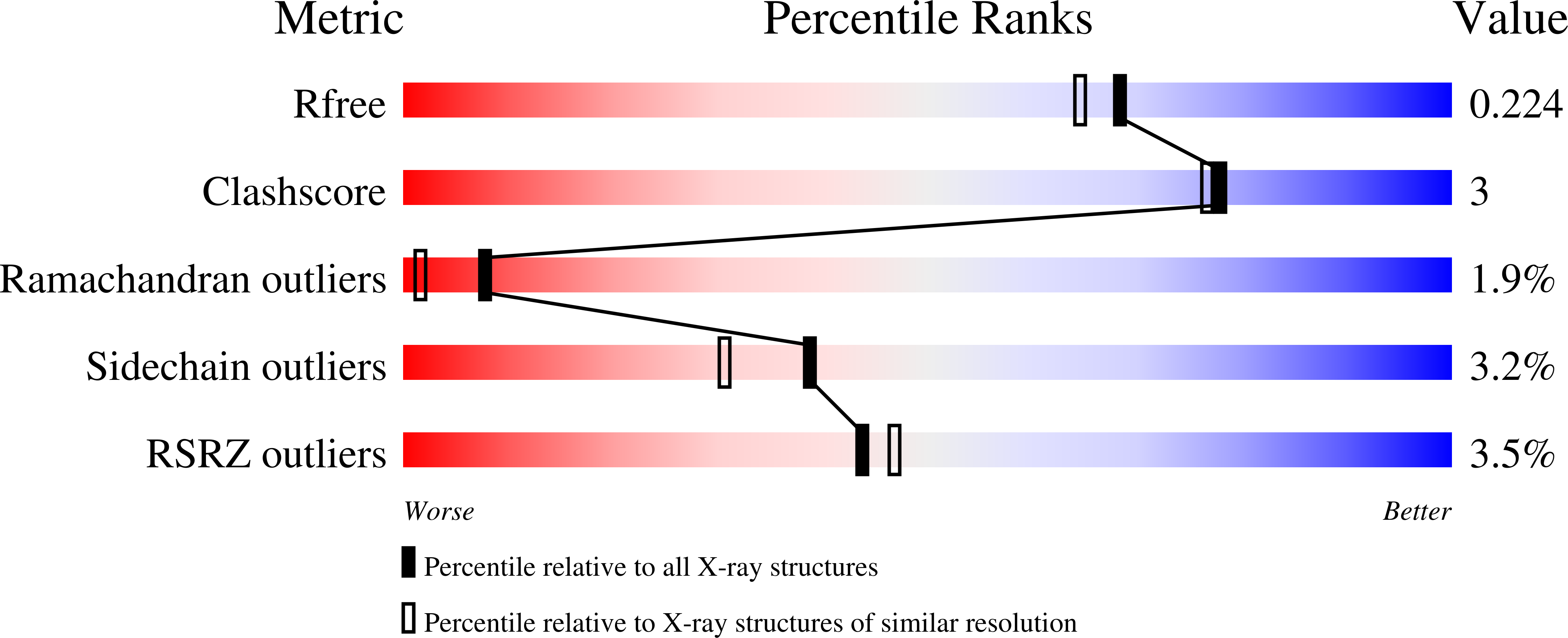
Resolution:
1.90 Å
R-Value Free:
0.23
R-Value Work:
0.20
R-Value Observed:
0.20
Space Group:
P 61 2 2
Macromolecular Entities
Polymer Type:polypeptide(L)
Molecule:Transcriptional activator cadC
Gene (Uniprot):cadC
Mutations:D471E
Chain IDs:A
Chain Length:372
Number of Molecules:1
Biological Source:Escherichia coli
Primary Citation
Crystal structure of the sensory domain of Escherichia coli CadC, a member of the ToxR-like protein family.
Protein Sci. 20 656 669 (2011)
PMID: 21308846 DOI: 10.1002/pro.594

Abstact

The membrane-integral transcriptional activator CadC comprises sensory and transcriptional regulatory functions within one polypeptide chain. Its C-terminal periplasmic domain, CadC(pd), is responsible for sensing of environmental pH as well as for binding of the feedback inhibitor cadaverine. Here we describe the crystal structure of CadC(pd) (residues 188-512) solved at a resolution of 1.8 Å via multiple wavelength anomalous dispersion (MAD) using a ReCl(6)(2-) derivative. CadC(pd) reveals a novel fold comprising two subdomains: an N-terminal subdomain dominated by a β-sheet in contact with three α-helices and a C-terminal subdomain formed by an eleven-membered α-helical bundle, which is oriented almost perpendicular to the helices in the first subdomain. Further to the native protein, crystal structures were also solved for its variants D471N and D471E, which show functionally different behavior in pH sensing. Interestingly, in the heavy metal derivative of CadC(pd) used for MAD phasing a ReCl(6)(2-) ion was found in a cavity located between the two subdomains. Amino acid side chains that coordinate this complex ion are conserved in CadC homologues from various bacterial species, suggesting a function of the cavity in the binding of cadaverine, which was supported by docking studies. Notably, CadC(pd) forms a homo-dimer in solution, which can be explained by an extended, albeit rather polar interface between two symmetry-related monomers in the crystal structure. The occurrence of several acidic residues in this region suggests protonation-dependent changes in the mode of dimerization, which could eventually trigger transcriptional activation by CadC in the bacterial cytoplasm.

Legend

Protein

Chemical

Disease

Primary Citation of related structures