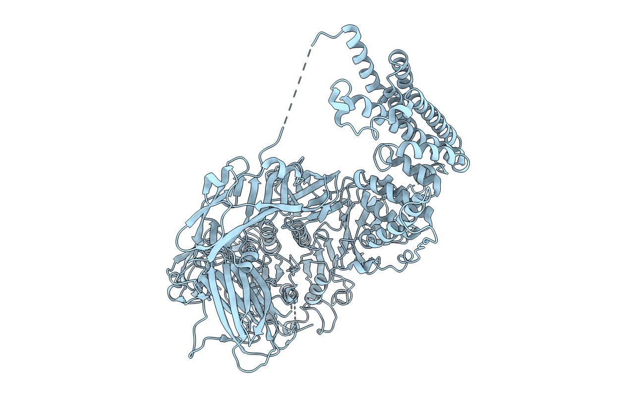
3LXU image
Entry Detail
PDB ID:
3LXU
Keywords:
Title:
Crystal Structure of Tripeptidyl Peptidase 2 (TPP II)
Biological Source:
Source Organism:
Host Organism:
PDB Version:
Deposition Date:
2010-02-25
Release Date:
2010-08-11
Method Details:
Experimental Method:
Resolution:
3.14 Å
R-Value Free:
0.29
R-Value Work:
0.24
Space Group:
P 41 21 2
Macromolecular Entities
Polymer Type:polypeptide(L)
Description:Tripeptidyl-peptidase 2
Chain IDs:A (auth: X)
Chain Length:1354
Number of Molecules:1
Biological Source:Drosophila melanogaster
Modified Residue
Compound ID Chain ID Parent Comp ID Details 2D Image
MSE A MET SELENOMETHIONINE
Ligand Molecules
Primary Citation
Hybrid molecular structure of the giant protease tripeptidyl peptidase II.
Nat.Struct.Mol.Biol. 17 990 996 (2010)
PMID: 20676100 DOI: 10.1038/nsmb.1870

Abstact

Tripeptidyl peptidase II (TPP II) is the largest known eukaryotic protease (6 MDa). It is believed to act downstream of the 26S proteasome, cleaving tripeptides from the N termini of longer peptides, and it is implicated in numerous cellular processes. Here we report the structure of Drosophila TPP II determined by a hybrid approach. We solved the structure of the dimer by X-ray crystallography and docked it into the three-dimensional map of the holocomplex, which we obtained by single-particle cryo-electron microscopy. The resulting structure reveals the compartmentalization of the active sites inside a system of chambers and suggests the existence of a molecular ruler determining the size of the cleavage products. Furthermore, the structure suggests a model for activation of TPP II involving the relocation of a flexible loop and a repositioning of the active-site serine, coupling it to holocomplex assembly and active-site sequestration.

Legend

Protein

Chemical

Disease

Primary Citation of related structures