3LV3 image
Deposition Date 2010-02-19
Release Date 2010-11-24
Last Version Date 2024-10-09
Entry Detail
PDB ID:
3LV3
Keywords:
Title:
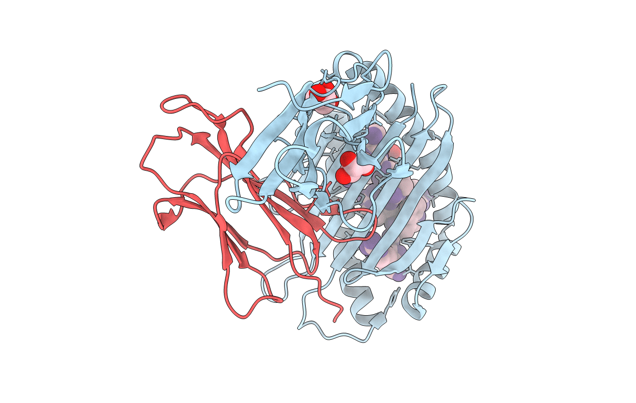
Crystal structure of HLA-B*2705 complexed with a peptide derived from the human voltage-dependent calcium channel alpha1 subunit (residues 513-521)
Biological Source:
Source Organism:
Homo sapiens (Taxon ID: 9606)
Host Organism:
Method Details:
Experimental Method:
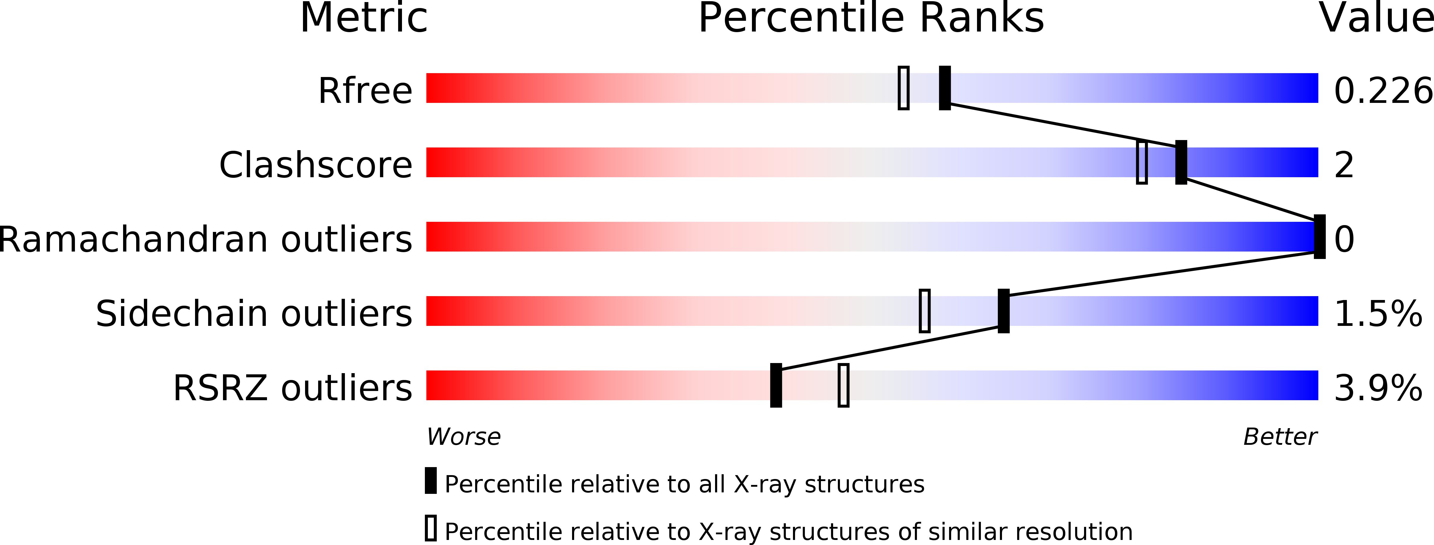
Resolution:
1.94 Å
R-Value Free:
0.22
R-Value Work:
0.17
R-Value Observed:
0.17
Space Group:
P 1 21 1
Macromolecular Entities
Polymer Type:polypeptide(L)
Molecule:HLA class I histocompatibility antigen, B-27 alpha chain
Chain IDs:A
Chain Length:276
Number of Molecules:1
Biological Source:Homo sapiens
Polymer Type:polypeptide(L)
Molecule:Beta-2-microglobulin
Gene (Uniprot):B2M
Chain IDs:B
Chain Length:100
Number of Molecules:1
Biological Source:Homo sapiens
Polymer Type:polypeptide(L)
Molecule:9-meric peptide from Voltage-dependent L-type calcium channel subunit alpha-1D
Gene (Uniprot):CACNA1D
Chain IDs:C
Chain Length:9
Number of Molecules:1
Biological Source:Homo sapiens
Ligand Molecules
Primary Citation
Loss of recognition by cross-reactive T cells and its relation to a C-terminus-induced conformational reorientation of an HLA-B*2705-bound peptide.
Protein Sci. 20 278 290 (2011)
PMID: 21280120 DOI: 10.1002/pro.559

Abstact

The human major histocompatibility complex class I antigen HLA-B*2705 binds several sequence-related peptides (pVIPR, RRKWRRWHL; pLPM2, RRRWRRLTV; pGR, RRRWHRWRL). Cross-reactivity of cytotoxic T cells (CTL) against these HLA-B*2705:peptide complexes seemed to depend on a particular peptide conformation that is facilitated by the engagement of a crucial residue within the binding groove (Asp116), associated with a noncanonical bulging-in of the middle portion of the bound peptide. We were interested whether a conformational reorientation of the ligand might contribute to the lack of cross-reactivity of these CTL with a peptide derived from voltage-dependent calcium channel α1 subunit (pCAC, SRRWRRWNR), in which the C-terminal peptide residue pArg9 could engage Asp116. Analyses of the HLA-B*2705:pCAC complex by X-ray crystallography at 1.94 Å resolution demonstrated that the peptide had indeed undergone a drastic reorientation, leading it to adopt a canonical binding mode accompanied by the loss of molecular mimicry between pCAC and sequence-related peptides such as pVIPR, pLMP2, and pGR. This was clearly a consequence of interactions of pArg9 with Asp116 and other F-pocket residues. Furthermore, we observed an unprecedented reorientation of several additional residues of the HLA-B*2705 heavy chain near the N-terminal region of the peptide, including also the presence of double conformations of two glutamate residues, Glu63 and Glu163, on opposing sides of the peptide binding groove. Together with the Arg-Ser exchange at peptide position 1, there are thus multiple structural reasons that may explain the observed failure of pVIPR-directed, HLA-B*2705-restricted CTL to cross-react with HLA-B*2705:pCAC complexes.

Legend

Protein

Chemical

Disease

Primary Citation of related structures