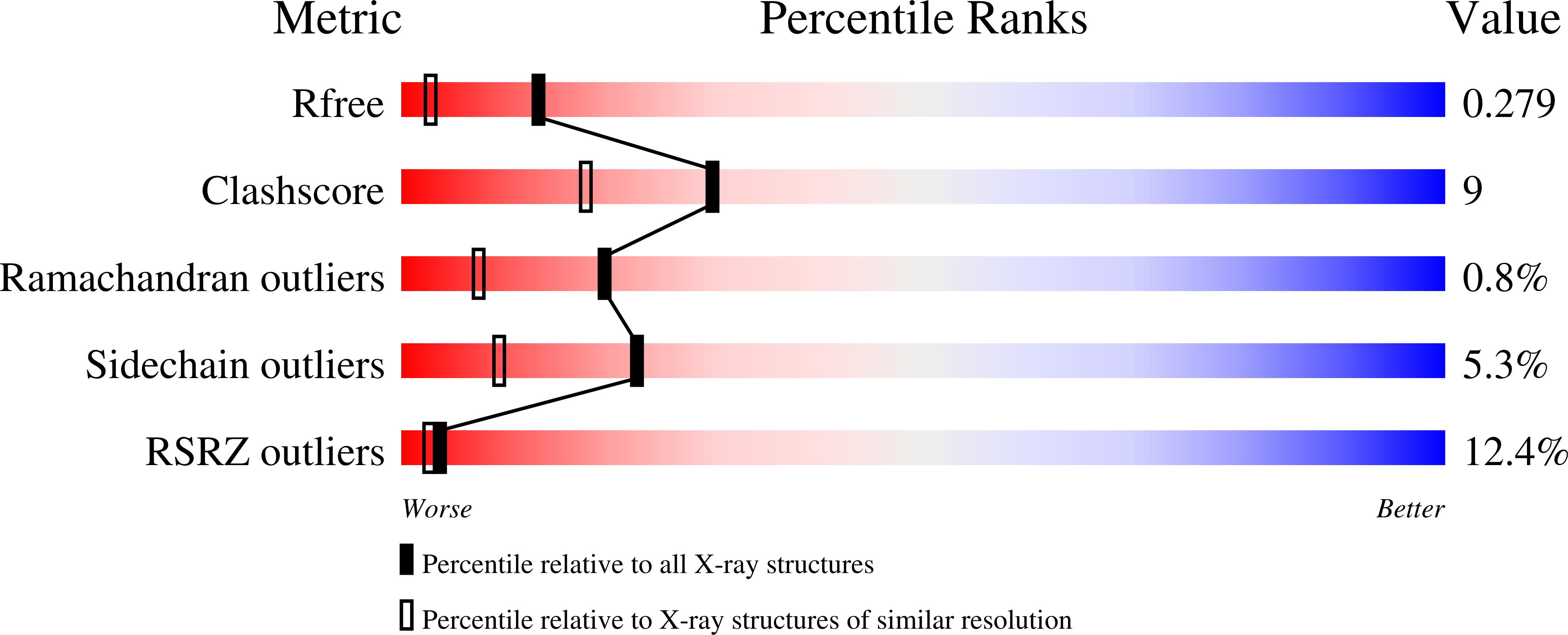
3LU9 image
Deposition Date 2010-02-17
Release Date 2010-03-16
Last Version Date 2024-11-06
Entry Detail
PDB ID:
3LU9
Keywords:
Title:
Crystal structure of human thrombin mutant S195A in complex with the extracellular fragment of human PAR1
Biological Source:
Source Organism:
Homo sapiens (Taxon ID: 9606)
Host Organism:
Method Details:
Experimental Method:
Resolution:
1.80 Å
R-Value Free:
0.23
R-Value Work:
0.19
R-Value Observed:
0.19
Space Group:
P 1
Macromolecular Entities
Polymer Type:polypeptide(L)
Molecule:Prothrombin
Gene (Uniprot):F2
Mutations:S195A
Chain IDs:A, D
Chain Length:46
Number of Molecules:2
Biological Source:Homo sapiens
Polymer Type:polypeptide(L)
Molecule:Prothrombin
Gene (Uniprot):F2
Chain IDs:B, E
Chain Length:259
Number of Molecules:2
Biological Source:Homo sapiens
Polymer Type:polypeptide(L)
Molecule:Proteinase-activated receptor 1
Gene (Uniprot):F2R
Chain IDs:C, F
Chain Length:25
Number of Molecules:2
Biological Source:Homo sapiens
Primary Citation
Crystal structure of thrombin bound to the uncleaved extracellular fragment of PAR1.
J.Biol.Chem. 285 15393 15398 (2010)
PMID: 20236938 DOI: 10.1074/jbc.M110.115337

Abstact

Abundant structural information exists on how thrombin recognizes ligands at the active site or at exosites separate from the active site region, but remarkably little is known about how thrombin recognizes substrates that bridge both the active site and exosite I. The case of the protease-activated receptor PAR1 is particularly relevant in view of the plethora of biological effects associated with its activation by thrombin. Here, we present the 1.8 A resolution structure of thrombin S195A in complex with a 30-residue long uncleaved extracellular fragment of PAR1 that documents for the first time a productive binding mode bridging the active site and exosite I. The structure reveals two unexpected features of the thrombin-PAR1 interaction. The acidic P3 residue of PAR1, Asp(39), does not hinder binding to the active site and actually makes favorable interactions with Gly(219) of thrombin. The tethered ligand domain shows a considerable degree of disorder even when bound to thrombin. The results fill a significant gap in our understanding of the molecular mechanisms of recognition by thrombin in ways that are relevant to other physiological substrates.

Legend

Protein

Chemical

Disease

Primary Citation of related structures