3LTG image
Deposition Date 2010-02-15
Release Date 2010-08-25
Last Version Date 2024-10-30
Entry Detail
PDB ID:
3LTG
Title:
Crystal structure of the Drosophila Epidermal Growth Factor Receptor ectodomain complexed with a low affinity Spitz mutant
Biological Source:
Source Organism:
Host Organism:
Method Details:
Experimental Method:
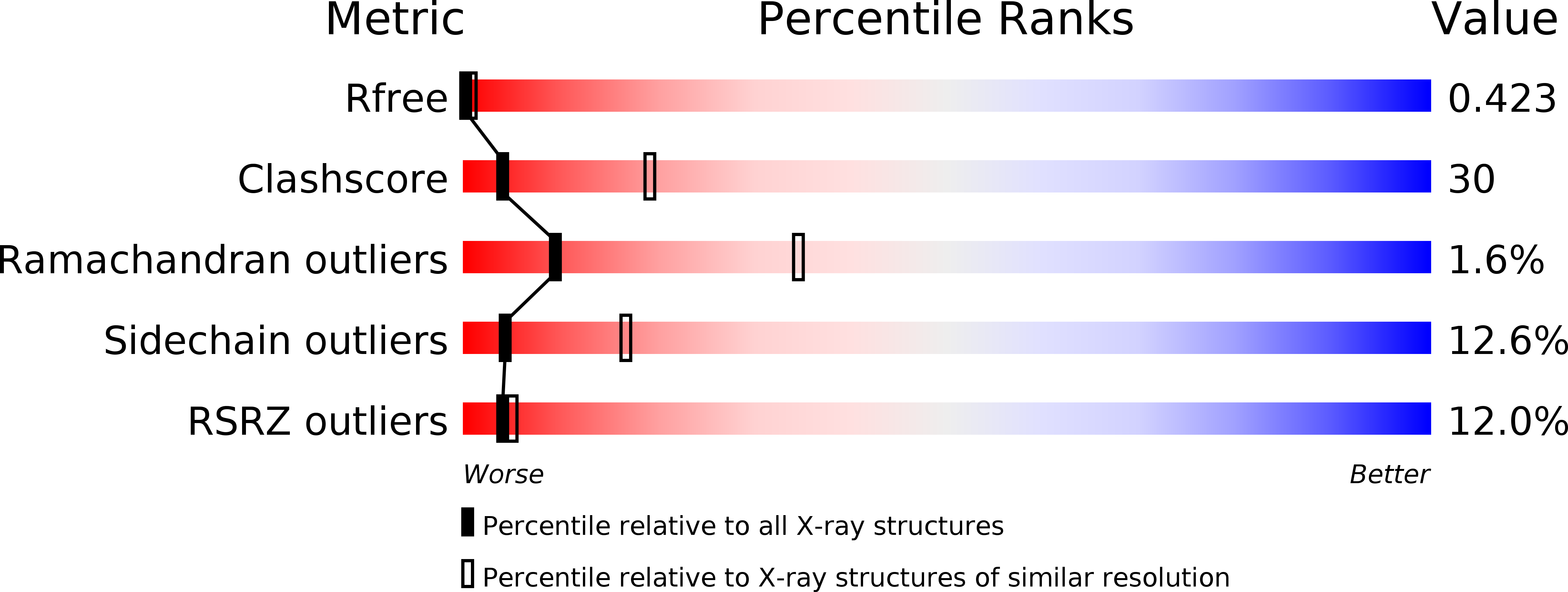
Resolution:
3.40 Å
R-Value Free:
0.42
R-Value Work:
0.40
R-Value Observed:
0.40
Space Group:
P 21 21 21
Macromolecular Entities
Structures with similar UniProt ID
Protein Blast
Polymer Type:polypeptide(L)
Molecule:Epidermal growth factor receptor
Gene (Uniprot):Egfr
Chain IDs:A, B (auth: C)
Chain Length:601
Number of Molecules:2
Biological Source:Drosophila melanogaster
Structures with similar UniProt ID
Protein Blast
Polymer Type:polypeptide(L)
Molecule:Protein spitz
Gene (Uniprot):spi
Chain IDs:C (auth: D)
Chain Length:52
Number of Molecules:1
Biological Source:Drosophila melanogaster
Ligand Molecules
Primary Citation
Structural basis for negative cooperativity in growth factor binding to an EGF receptor.
Cell(Cambridge,Mass.) 142 568 579 (2010)
PMID: 20723758 DOI: 10.1016/j.cell.2010.07.015

Abstact

Transmembrane signaling by the epidermal growth factor receptor (EGFR) involves ligand-induced dimerization and allosteric regulation of the intracellular tyrosine kinase domain. Crystallographic studies have shown how ligand binding induces dimerization of the EGFR extracellular region but cannot explain the "high-affinity" and "low-affinity" classes of cell-surface EGF-binding sites inferred from curved Scatchard plots. From a series of crystal structures of the Drosophila EGFR extracellular region, we show here how Scatchard plot curvature arises from negatively cooperative ligand binding. The first ligand-binding event induces formation of an asymmetric dimer with only one bound ligand. The unoccupied site in this dimer is structurally restrained, leading to reduced affinity for binding of the second ligand, and thus negative cooperativity. Our results explain the cell-surface binding characteristics of EGF receptors and suggest how individual EGFR ligands might stabilize distinct dimeric species with different signaling properties.

Legend

Protein

Chemical

Disease

Primary Citation of related structures
Feedback Form
Name
Email
Institute
Feedback