3LS0 image
Deposition Date 2010-02-12
Release Date 2010-03-31
Last Version Date 2024-03-20
Entry Detail
PDB ID:
3LS0
Keywords:
Title:
Crystal Structure of Cyanobacterial PsbQ from Synechocystis sp. PCC 6803
Biological Source:
Source Organism:
Host Organism:
Method Details:
Experimental Method:
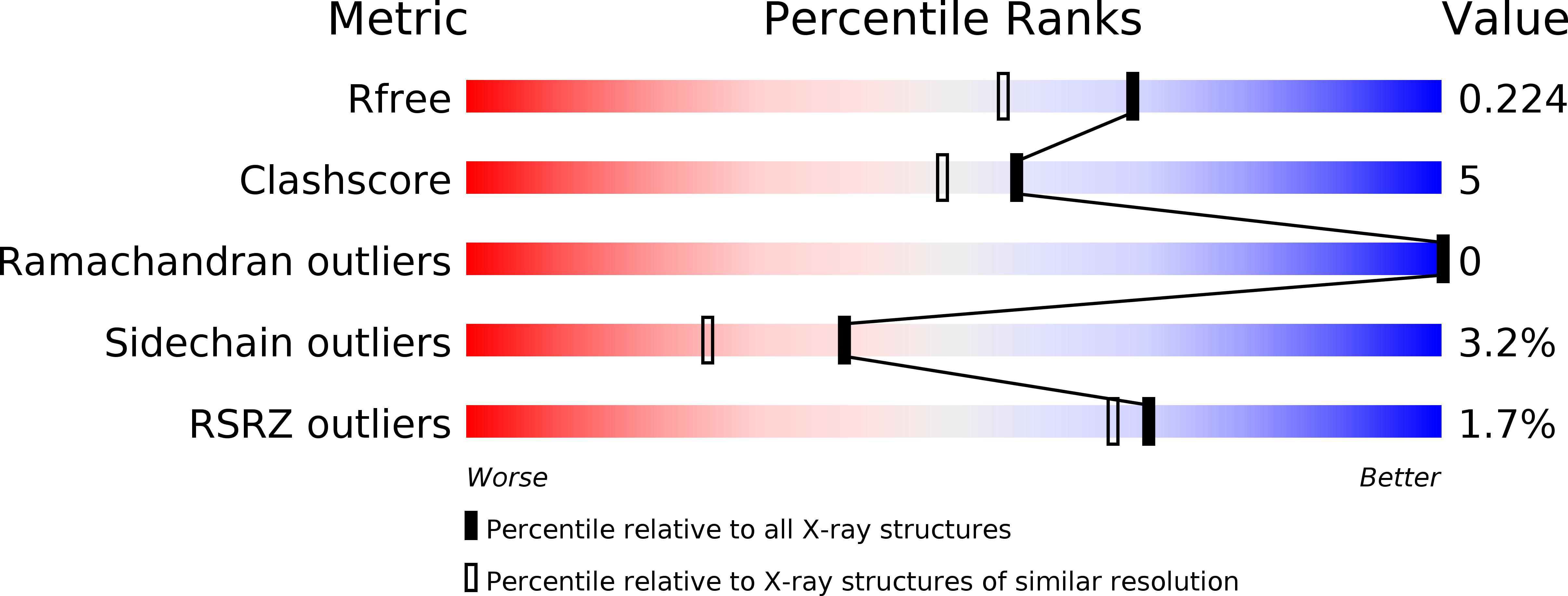
Resolution:
1.80 Å
R-Value Free:
0.20
R-Value Work:
0.16
R-Value Observed:
0.16
Space Group:
P 21 21 21
Macromolecular Entities
Structures with similar UniProt ID
Protein Blast
Polymer Type:polypeptide(L)
Molecule:Sll1638 protein
Gene (Uniprot):psbQ
Chain IDs:A
Chain Length:133
Number of Molecules:1
Biological Source:Synechocystis sp.
Primary Citation
Crystal Structure of PsbQ from Synechocystis sp. PCC 6803 at 1.8 A: Implications for Binding and Function in Cyanobacterial Photosystem II
Biochemistry 49 2765 2767 (2010)
PMID: 20210304 DOI: 10.1021/bi100217h

Abstact

In Synechocystis sp. PCC 6803, PsbQ is associated with photosystem II (PSII) complexes with the highest activity and stability. However, this subunit is not found in PSII X-ray crystallographic structures from Thermosynechococcus elongatus or Thermosynechococcus vulcanus. We present the crystal structure of cyanobacterial PsbQ determined in the presence and absence of Zn(2+). The protein has a well-defined helical core, containing four helices arranged in an up-down-up-down fold. A conserved potential interaction site composed of a divalent metal binding site and adjacent hydrophobic pocket has been identified.

Legend

Protein

Chemical

Disease

Primary Citation of related structures