3LRH image
Deposition Date 2010-02-11
Release Date 2011-02-23
Last Version Date 2023-09-06
Entry Detail
PDB ID:
3LRH
Keywords:
Title:
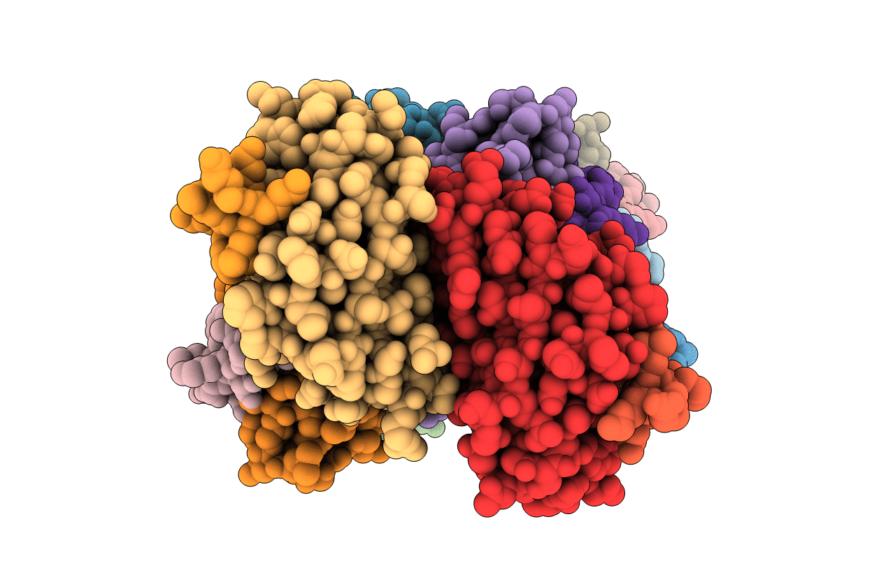
Structure of anti-huntingtin VL domain in complex with huntingtin peptide
Biological Source:
Source Organism:
Homo sapiens (Taxon ID: 9606)
Host Organism:
Method Details:
Experimental Method:
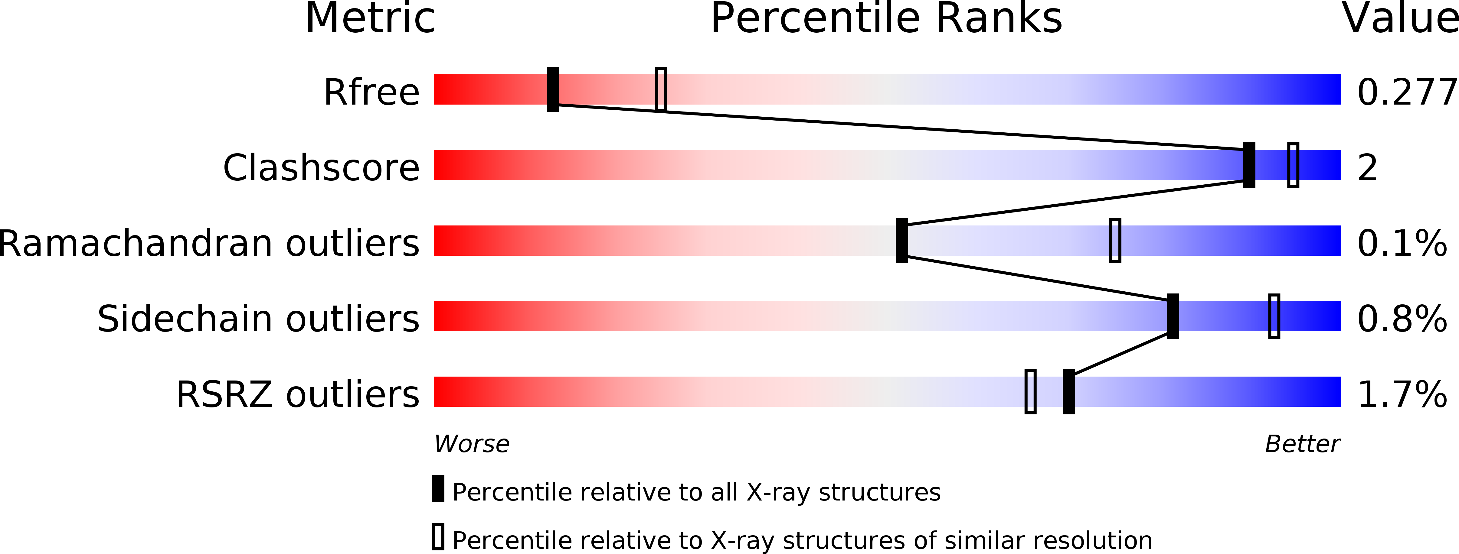
Resolution:
2.60 Å
R-Value Free:
0.27
R-Value Work:
0.21
R-Value Observed:
0.21
Space Group:
P 1 21 1
Macromolecular Entities
Polymer Type:polypeptide(L)
Molecule:anti-huntingtin VL domain
Mutagens:G112A
Chain IDs:A, C, E, G, I, K, M, O
Chain Length:121
Number of Molecules:8
Biological Source:Homo sapiens
Structures with similar UniProt ID
Protein Blast
Polymer Type:polypeptide(L)
Molecule:Huntingtin
Gene (Uniprot):HTT
Chain IDs:B, D, F, H, J, L, N, P
Chain Length:14
Number of Molecules:8
Biological Source:
Primary Citation
A Disulfide-Free Single-Domain V(L) Intrabody with Blocking Activity towards Huntingtin Reveals a Novel Mode of Epitope Recognition.
J.Mol.Biol. 414 337 355 (2011)
PMID: 21968397 DOI: 10.1016/j.jmb.2011.09.034

Abstact

We present the crystal structure and biophysical characterization of a human V(L) [variable domain immunoglobulin (Ig) light chain] single-domain intrabody that binds to the huntingtin (Htt) protein and has been engineered for antigen recognition in the absence of its intradomain disulfide bond, otherwise conserved in the Ig fold. Analytical ultracentrifugation demonstrated that the αHtt-V(L) 12.3 domain is a stable monomer under physiological conditions even at concentrations >20 μM. Using peptide SPOT arrays, we identified the minimal binding epitope to be EKLMKAFESLKSFQ, comprising the N-terminal residues 5-18 of Htt and including the first residue of the poly-Gln stretch. X-ray structural analysis of αHtt-V(L) both as apo protein and in the presence of the epitope peptide revealed several interesting insights: first, the role of mutations acquired during the combinatorial selection process of the αHtt-V(L) 12.3 domain-initially starting from a single-chain Fv fragment-that are responsible for its stability as an individually soluble Ig domain, also lacking the disulfide bridge, and second, a previously unknown mode of antigen recognition, revealing a novel paratope. The Htt epitope peptide adopts a purely α-helical structure in the complex with αHtt-V(L) and is bound at the base of the complementarity-determining regions (CDRs) at the concave β-sheet that normally gives rise to the interface between the V(L) domain and its paired V(H) (variable domain Ig heavy chain) domain, while only few interactions with CDR-L1 and CDR-L3 are formed. Notably, this noncanonical mode of antigen binding may occur more widely in the area of in vitro selected antibody fragments, including other Ig-like scaffolds, possibly even if a V(H) domain is present.

Legend

Protein

Chemical

Disease

Primary Citation of related structures