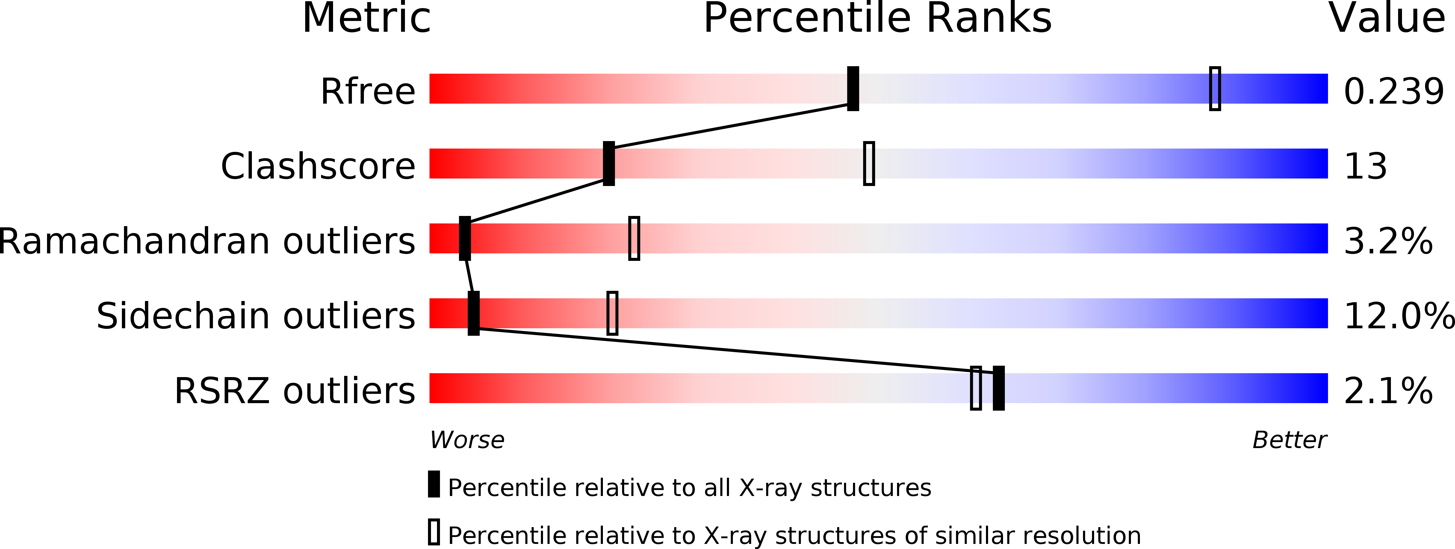
3LQZ image
Entry Detail
PDB ID:
3LQZ
Keywords:
Title:
Crystal Structure of HLA-DP2
Biological Source:
Source Organism:
PDB Version:
Deposition Date:
2010-02-10
Release Date:
2010-04-14
Method Details:
Experimental Method:
Resolution:
3.25 Å
R-Value Free:
0.25
R-Value Work:
0.21
R-Value Observed:
0.21
Space Group:
P 31 2 1
Macromolecular Entities
Polymer Type:polypeptide(L)
Description:HLA class II histocompatibility antigen, DP alpha 1 chain
Chain IDs:A
Chain Length:181
Number of Molecules:1
Biological Source:Homo sapiens
Polymer Type:polypeptide(L)
Description:HLA-DP2 beta chain linked with DRa peptide
Chain IDs:B
Chain Length:212
Number of Molecules:1
Biological Source:Homo sapiens
Modified Residue
Compound ID Chain ID Parent Comp ID Details 2D Image
ASN B ASN GLYCOSYLATION SITE
Ligand Molecules
Primary Citation
Crystal structure of HLA-DP2 and implications for chronic beryllium disease.
Proc.Natl.Acad.Sci.USA 107 7425 7430 (2010)
PMID: 20356827 DOI: 10.1073/pnas.1001772107

Abstact

Chronic beryllium disease (CBD) is a fibrotic lung disorder caused by beryllium (Be) exposure and is characterized by granulomatous inflammation and the accumulation of Be-responsive CD4(+) T cells in the lung. Genetic susceptibility to CBD has been associated with certain alleles of the MHCII molecule HLA-DP, especially HLA-DPB1*0201 and other alleles that contain a glutamic acid residue at position 69 of the beta-chain (betaGlu69). The HLA-DP alleles that can present Be to T cells match those implicated in the genetic susceptibility, suggesting that the HLA contribution to disease is based on the ability of those molecules to bind and present Be to T cells. The structure of HLA-DP2 and its interaction with Be are unknown. Here, we present the HLA-DP2 structure with its antigen-binding groove occupied by a self-peptide derived from the HLA-DR alpha-chain. The most striking feature of the structure is an unusual solvent exposed acidic pocket formed between the peptide backbone and the HLA-DP2 beta-chain alpha-helix and containing three glutamic acids from the beta-chain, including betaGlu69. In the crystal packing, this pocket has been filled with the guanidinium group of an arginine from a neighboring molecule. This positively charged moiety forms an extensive H-bond/salt bridge network with the three glutamic acids, offering a plausible model for how Be-containing complexes might occupy this site. This idea is strengthened by the demonstration that mutation of any of the three glutamic acids in this pocket results in loss of the ability of DP2 to present Be to T cells.

Legend

Protein

Chemical

Disease

Primary Citation of related structures