Deposition Date
2010-02-02
Release Date
2010-03-09
Last Version Date
2024-11-06
Entry Detail
PDB ID:
3LNJ
Keywords:
Title:
Crystal structure of human MDM2 in complex with D-peptide inhibitor (DPMI-alpha)
Method Details:
Experimental Method:
Resolution:
2.40 Å
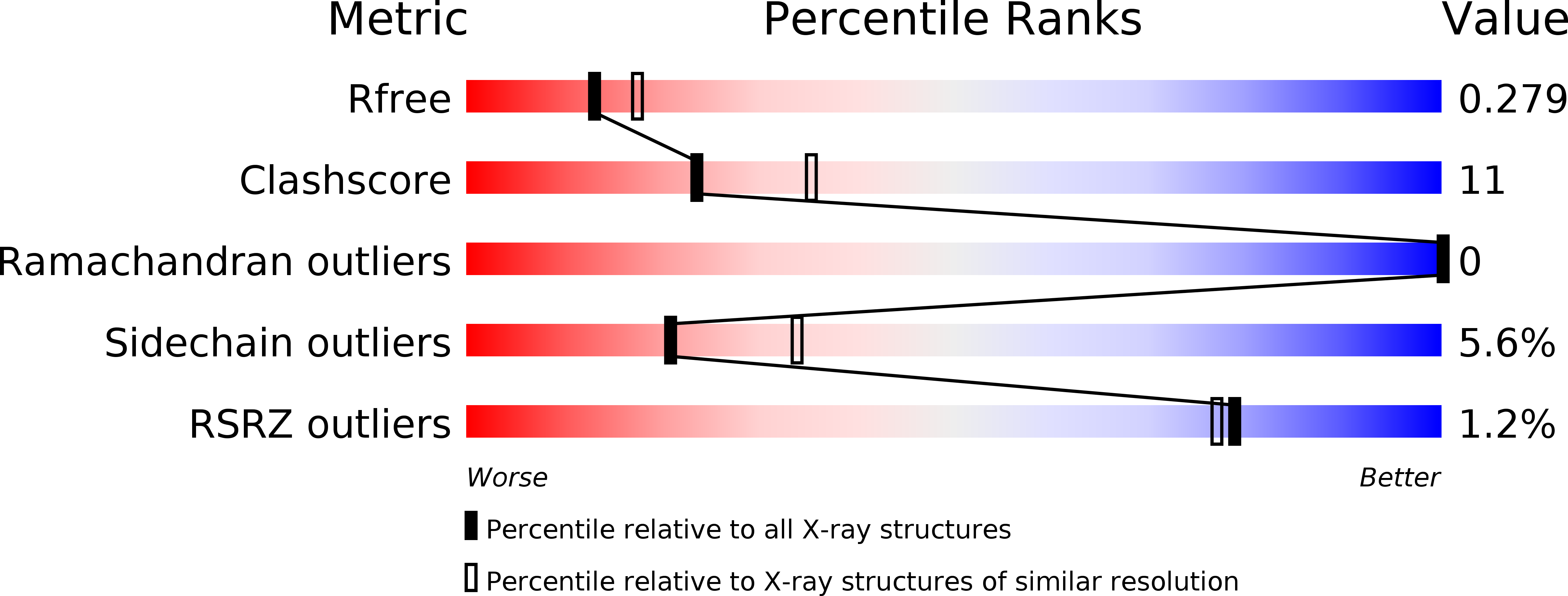
R-Value Free:
0.25
R-Value Work:
0.20
R-Value Observed:
0.21
Space Group:
C 2 2 21