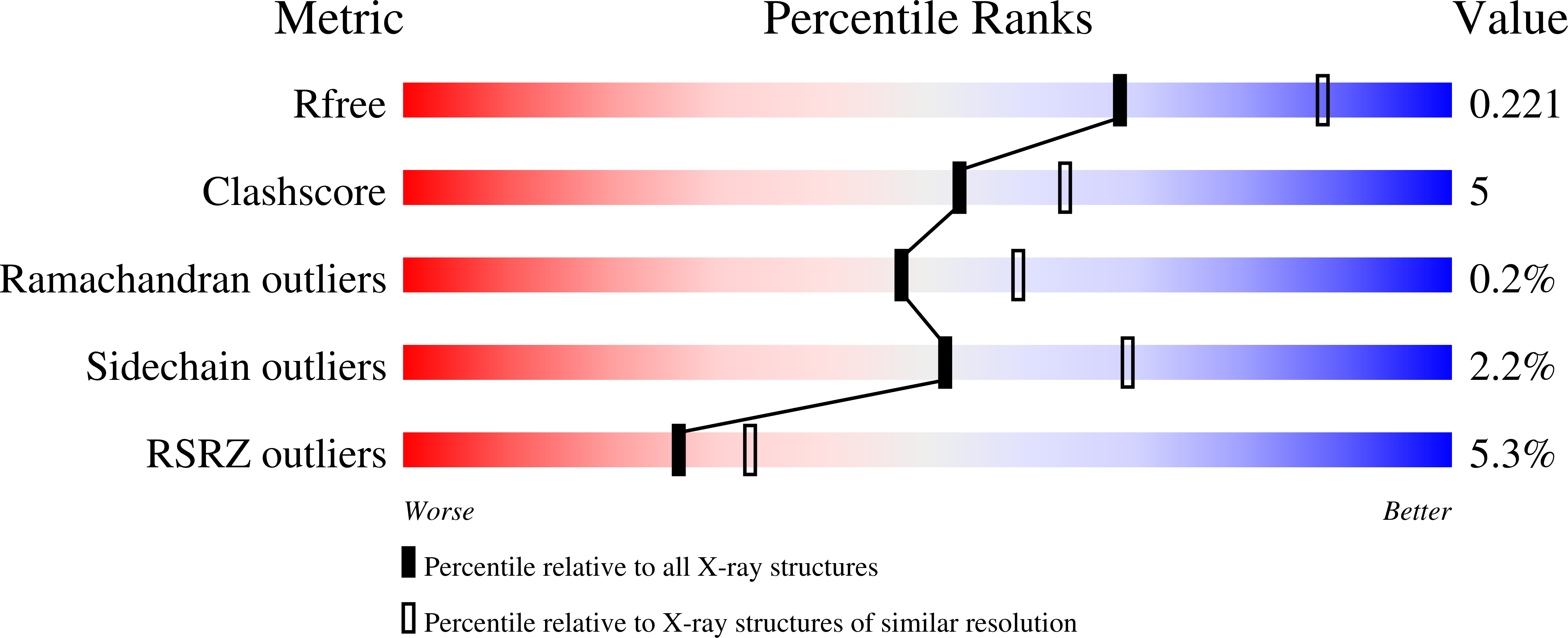
3LNI image
Deposition Date 2010-02-02
Release Date 2010-03-02
Last Version Date 2023-09-06
Entry Detail
PDB ID:
3LNI
Keywords:
Title:
Crystal structure of E-cadherin EC12 E89A
Biological Source:
Source Organism:
Mus musculus (Taxon ID: 10090)
Host Organism:
Method Details:
Experimental Method:
Resolution:
2.30 Å
R-Value Free:
0.22
R-Value Work:
0.16
R-Value Observed:
0.16
Space Group:
C 1 2 1
Macromolecular Entities
Polymer Type:polypeptide(L)
Molecule:Cadherin-1
Gene (Uniprot):Cdh1
Mutations:E89A
Chain IDs:A, B
Chain Length:213
Number of Molecules:2
Biological Source:Mus musculus
Ligand Molecules
Primary Citation
Two-step adhesive binding by classical cadherins.
Nat.Struct.Mol.Biol. 17 348 357 (2010)
PMID: 20190754 DOI: 10.1038/nsmb.1784

Abstact

Crystal structures of classical cadherins have revealed two dimeric configurations. In the first, N-terminal beta-strands of EC1 domains 'swap' between partner molecules. The second configuration (the 'X dimer'), also observed for T-cadherin, is mediated by residues near the EC1-EC2 calcium binding sites, and N-terminal beta-strands of partner EC1 domains, though held adjacent, do not swap. Here we show that strand-swapping mutants of type I and II classical cadherins form X dimers. Mutant cadherins impaired for X-dimer formation show no binding in short-time frame surface plasmon resonance assays, but in long-time frame experiments, they have homophilic binding affinities close to that of wild type. Further experiments show that exchange between monomers and dimers is slowed in these mutants. These results reconcile apparently disparate results from prior structural studies and suggest that X dimers are binding intermediates that facilitate the formation of strand-swapped dimers.

Legend

Protein

Chemical

Disease

Primary Citation of related structures