
Deposition Date
2010-02-02
Release Date
2011-01-26
Last Version Date
2023-11-01
Entry Detail
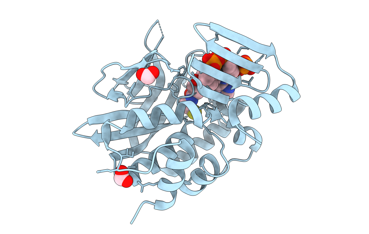
PDB ID:
3LNB
Keywords:
Title:
Crystal Structure Analysis of Arylamine N-acetyltransferase C from Bacillus anthracis
Biological Source:
Source Organism(s):
Bacillus anthracis (Taxon ID: 1392)
Expression System(s):
Method Details:
Experimental Method:
Resolution:
2.01 Å
R-Value Free:
0.24
R-Value Work:
0.20
Space Group:
P 41 21 2
