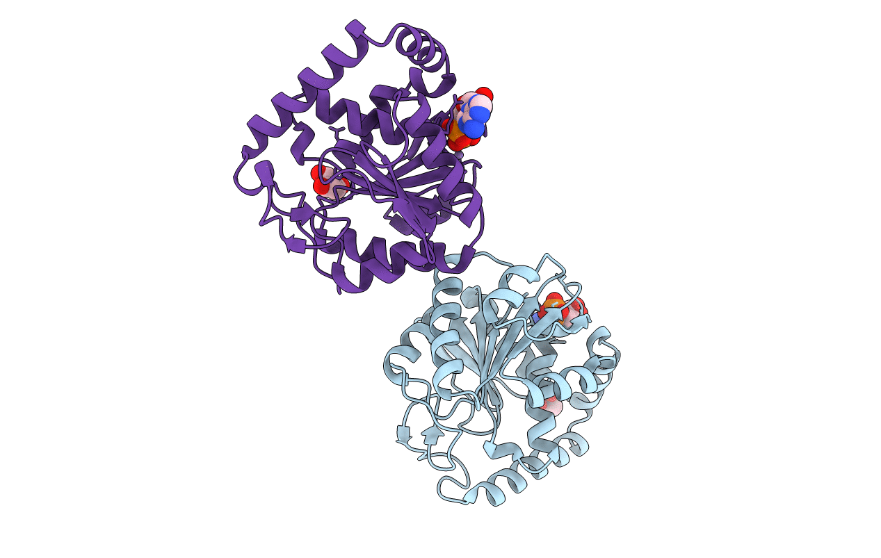
3LLM image
Entry Detail
PDB ID:
3LLM
Keywords:
Title:
Crystal Structure Analysis of a RNA Helicase
Biological Source:
Source Organism:
Host Organism:
PDB Version:
Deposition Date:
2010-01-29
Release Date:
2010-05-12
Method Details:
Experimental Method:
Resolution:
2.80 Å
R-Value Free:
0.23
R-Value Work:
0.21
R-Value Observed:
0.21
Space Group:
P 32 2 1
Macromolecular Entities
Polymer Type:polypeptide(L)
Description:ATP-dependent RNA helicase A
Chain IDs:A, B
Chain Length:235
Number of Molecules:2
Biological Source:Homo sapiens
Modified Residue
Compound ID Chain ID Parent Comp ID Details 2D Image
MSE A MET SELENOMETHIONINE
Primary Citation
Crystal structure of human RNA helicase A (DHX9): structural basis for unselective nucleotide base binding in a DEAD-box variant protein.
J.Mol.Biol. 400 768 782 (2010)
PMID: 20510246 DOI: 10.1016/j.jmb.2010.05.046

Abstact

RNA helicases of the DExD/H-box superfamily are critically involved in all RNA-related processes. No crystal structures of human DExH-box domains had been determined previously, and their structures were difficult to predict owing to the low level of homology among DExH-motif-containing proteins from diverse species. Here we present the crystal structures of the conserved domain 1 of the DEIH-motif-containing helicase DHX9 and of the DEAD-box helicase DDX20. Both contain a RecA-like core, but DHX9 differs from DEAD-box proteins in the arrangement of secondary structural elements and is more similar to viral helicases such as NS3. The N-terminus of the DHX9 core contains two long alpha-helices that reside on the surface of the core without contributing to nucleotide binding. The RNA-polymerase-II-interacting minimal transactivation domain sequence forms an extended loop structure that resides in a hydrophobic groove on the surface of the DEIH domain. DHX9 lacks base-selective contacts and forms an unspecific but important stacking interaction with the base of the bound nucleotide, and our biochemical analysis confirms that the protein can hydrolyze ATP, guanosine 5'-triphosphate, cytidine 5'-triphosphate, and uridine 5'-triphosphate. Together, these findings allow the localization of functional motifs within the three-dimensional structure of a human DEIH helicase and show how these enzymes can bind nucleotide with high affinity in the absence of a Q-motif.

Legend

Protein

Chemical

Disease

Primary Citation of related structures